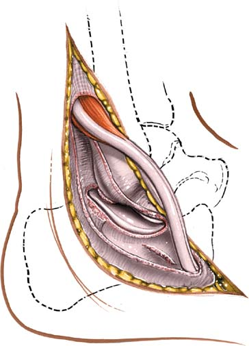
 Lateral Approach to the Posterior Talocalcaneal Joint
The lateral approach to the posterior talocalcaneal joint exposes the posterior facet of the talocalcaneal joint more extensively than does the anterolateral approach. It is mainly used for arthrodesis of the posterior part of the talocalcaneal joint.
Position of the Patient
Place the patient supine on the operating table with a sandbag under the buttock of the affected side to bring the lateral malleolus forward. Place a support on the opposite iliac crest, then tilt the table 20 degrees to 30 degrees away from the surgeon to improve access still further. Exsanguinate the limb either by elevating it for 3 to 5 minutes or by applying a soft rubber bandage, then inflate a tourniquet (see Fig. 7-1).
Landmarks and Incision
Landmarks
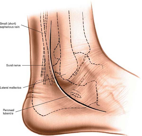
The lateral malleolus is the subcutaneous distal end of the fibula. The peroneal tubercle is a small protuberance of bone on the lateral surface of the calcaneus that separates the tendons of the peroneus longus and brevis muscles. It lies distal and anterior to the lateral malleolus.
Incision
Make a curved incision 10 to 13 cm long on the lateral aspect of the ankle. Begin some 4 cm above the tip of the lateral malleolus on the posterior border of the fibula. Follow the posterior border of the fibula down to the tip of the lateral malleolus, and then curve the incision forward, passing over the peroneal tubercle parallel to the course of the peroneal tendons (Fig. 12-1).
Internervous Plane
No internervous plane exists in this approach. The peroneus muscles, whose tendons are mobilized and retracted anteriorly, share a nerve supply from the superficial peroneal nerve. The approach is safe because the muscles receive their supply at a point well proximal to it.
Superficial Surgical Dissection
Stay updated, free articles. Join our Telegram channel

Full access? Get Clinical Tree


