 Lateral Approach to the Lateral Malleolus
The approach to the lateral malleolus is used primarily for open reduction and internal fixation of lateral malleolar fractures. It also offers access to the posterolateral aspect of the tibia.
Position of the Patient
Place the patient supine on the operating table with a sandbag under the buttock of the affected limb. The sandbag causes the limb to rotate medially, bringing the lateral malleolus forward and making it easier to reach (Fig. 7-1). Tilt the table away from you to further increase the internal rotation of the limb. Operating with the patient on his or her side also provides excellent access to the distal fibula, but the medial malleolus cannot be reached unless the patient’s position is changed, something that is necessary in the fixation of bimalleolar fractures (Fig. 7-2). Exsanguinate the limb by elevating it for 3 to 5 minutes, then inflate a tourniquet.
Landmarks and Incision
Landmarks
Palpate the subcutaneous surface of the fibula and the lateral malleolus, which lies at its distal end.
The short saphenous vein can be seen running along the posterior border of the lateral malleolus before the limb is exsanguinated.
Incision
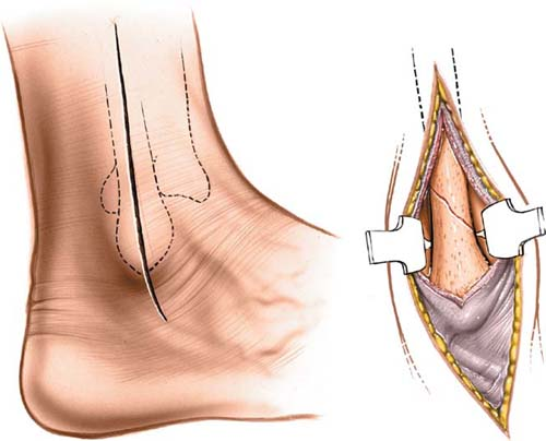
Make a 10- to 15-cm longitudinal incision along the posterior margin of the fibula all the way to its distal end and continuing for a further 2 cm (Fig. 7-3A). In fracture surgery, center the incision at the level of the fracture.
Internervous Plane
There is no internervous plane, because the dissection is being performed down to a subcutaneous bone. For higher fractures of the fibula, the internervous plane lies between the peroneus tertius muscle (which is supplied by the deep peroneal nerve) and the peroneus brevis muscle (which is supplied by the superficial peroneal nerve).1
Stay updated, free articles. Join our Telegram channel

Full access? Get Clinical Tree


