Knee
Acute Injuries
11.1 Anterior Cruciate Ligament (ACL)
Sports Med 2003;33:455; Am J Knee Surg 1998;11:128
Cause:
Mechanism of injury is direct blow to the lateral, medial, or anterior aspects of the knee causing valgus or varus strain, twisting (rotational), or hyperextension.
Epidem:
Varies by activity: alpine skiing, 7/1,000 skier days; collegiate football, 5/1,000 player days; general population, 3/1,000 knee injuries.
Women appear to have increased risk in some sports compared with men. Thought to be related to increased Q angle (formed by the line from the ASIS to the center of the patella, and a line from the center of the patella to the tibial tubercle), narrower femoral notch, and smaller diameter ligament.
Pathophys:
Major stabilizer preventing anterior translation of the tibia with respect to the femur, secondary rotational stabilizer.
Injuries to other intra-articular structures (menisci, collateral ligaments) are commonly associated.
Sx:
Patient typically describes a substantial trauma to the knee and often reports hearing or feeling a pop.
Followed (within 2 hr) by a massive joint effusion (hemarthrosis).
Pain is diffuse and typically related to bone bruising and/or meniscal injury.
In chronic injuries, often complain of instability with quick turns or pivot movements.
Si:
Acutely, substantial intra-articular effusion is palpable; joint line or posterior tenderness is common.
Patency of neurovascular structures should be assessed.
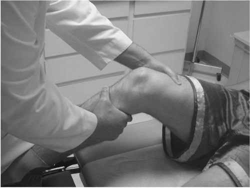
Lachman test: the patient is supine and relaxed, the knee is flexed to 20°, stabilize the distal femur with one hand while exerting an anteriorly directed force on the proximal tibia. Excessive anterior translation of the tibia when compared to the uninjured knee indicates laxity (Figure 11.1).
Pivot shift: the patient is supine and the leg extended with the foot internally rotated, a valgus force is applied at the knee as the knee is slowly flexed. At approximately 30° a rotatory clunk will indicate a pos test.
Diff Dx:
Crs:
Acute symptoms subside over 4-6 w; effusion may persist for 8-12 w.
Instability and re-injury of intra-articular structures common.
With recurrent instability, gradual degenerative disease is common.
X-ray:
Radiographs will occasionally demonstrate a Segond lesion (avulsion fracture at the lateral proximal tibia).
MRI is confirmatory, but not always necessary, however is useful to evaluate for other associated intra-articular injuries.
Rx:
Initial: knee immobilizer for 24-72 hr with crutch ambulation. PRICEMM is useful for symptom relief. Structured therapy program should be instituted early to limit strength loss and maintain range of motion.
Subacute: structured therapy including proprioception training and strengthening. Functional bracing for activities.
Surgery: indication for surgery is persistent instability. Determinates include: age, chosen sport, level of participation, response to therapy. Surgery is usually delayed 2-6 w after injury.
Return to Activity:
Nonoperative treatment, graded return to sport over 6-12 w.
Surgical treatment, return to play 6-12 months following surgery.
11.2 Posterior Cruciate Injuries
Am J Sports Med 2004;32:361; Am J Knee Surg 1996;9:200
Cause:
Epidem:
Much less common than ACL injuries.
Most common in football and hockey.
NFL draftees have approximately 2% incidence.
Pathophys:
Arises proximally on the medial femoral condyle in the intracondylar notch. The fibers blend with the posterior capsular fibers at insertion on the proximal posterior tibia.
The complex biomechanical architecture primarily functions to prevent posterior translation of the tibial with respect to the femur.
The PCL also limits hyperextension and internal rotation.
Sx:
Acute: pain in the posterior/lateral knee, swelling and occasionally instability with deceleration motions (as in walking down stairs).
Chronic instability.
Si:
Posterolateral tenderness, effusion. Positive instability testing.
Posterior drawer sign: with the patient supine, flex the hip to 45° and the knee to 90° with the foot flat on the table; a posteriorly directed force is applied to the proximal anterior tibia. Increased translation indicates PCL injury.
Sag sign: with the patient supine, the hips and knees are flexed to 90° with both feet supported above the table, a torn PCL will allow the tibial to translate posteriorly in this position in comparison to the uninjured knee.
Cmplc:
Chronic instability leads to DJD.
Instability can lead to damage to other intra-articular structures.
Diff Dx:
Crs: Instability may develop late (years after injury).
X-ray:
Radiographs usually negative.
MRI typically diagnostic.
Rx:
Initial and nonoperative treatment as for ACL injuries.
11.3 Meniscus Injuries
J Anat 1998;193:161; Am J Sports Med 2004;32:337
Cause:
Twisting injury with the foot planted, valgus or varus strain, hyperextension, or hyperflexion.
Often the trauma will appear minor or no specific event is recalled on history.
Epidem:
Approximately 60/100,000 general population.
Most common in cutting sports, football, soccer, basketball, wrestling.
Pathophys:
Two “C” shaped fibrocartilagenous structures anchored via the capsule to the tibial plateau.
They are thinner (and thus avascular) centrally, with the periphery relatively well vascularized.
Function in both load sharing to dissipate forces on the proximal tibia, and in maintaining joint integrity.
Sx:
An appropriate mechanism may be described, mild to moderate swelling, joint line pain, pain with flexion or extension, and a sensation of locking or catching within the knee joint.
Si:
Effusion, joint line tenderness, and painful passive range of motion. Positive meniscal signs:
The Mcmurrays test: with the patient supine the hip is flexed to 90° and the knee maximally flexed, the examiner then internally or externally rotates the tibia and extends the knee while exerting a valgus or varus force respectively at the knee. A Mcmurrays test is positive if these maneuvers produce a painful pop.
The Apley’s compression test: with the patient prone the knee is flexed to 90° and a load is applied to the tibia while the tibia is internally and externally rotated; this will cause pain with a damaged meniscus.
Complc:
Meniscal degeneration.
Degenerative joint disease.
Locked knee.
Diff Dx:
Other causes of joint swelling (fracture, ligament injury, DJD).
Other causes of locking or catching (patellar dysfunction).
Other medial or lateral pain entities (ITB friction syndrome, bursitis).
Crs: Acute symptoms (pain with weight bearing, swelling, painful ROM) last 2-3 w with gradual improvement over 8-12 w.
X-ray:
Plain radiographs are negative.
MRI scan is useful for confirming diagnosis, but unnecessary if clinical evaluation conclusive.
Rx:
Nonoperative treatment is effective in 50-75% of pts with an uncomplicated meniscal tear.
Initially, PRICEMM measures with activity modification including short-term crutch use and limited walking for 3-5 d will reduce pain symptoms.
Following the initial rest period, the patient should initiate exercise aimed at maintaining strength and flexibility while limiting pain. Alternate activities include cycling, walking, and pool running or swimming.
As symptoms resolve, a gradual running program can be instituted with low intensity, short runs without hills or turns. Cutting and twisting activities should be avoided for 8-10 w.
If symptoms fail to resolve in 6-12 w, the pt should be referred for diagnostic imaging (MRI) or surgical evaluation.
In a mechanically locked knee (ie, a loss of motion due to meniscal impingement) referral for urgent reduction or surgery should be arranged.
Return to Activity: As outlined above.
11.4 Patellar Dislocation and Subluxation
Acta Orthop Scand 1997;68:419
Cause:
The most common mechanism is a twisting or valgus motion with a forceful quadriceps contraction.
May be caused by direct blow to medial patella.
Epidem: Reported in a wide variety of sports. Most frequent in jumping and contact activities.
Pathophys:
The patella usually dislocates laterally, over the femoral condyle.
This displacement may be a true dislocation or may be partial which spontaneously reduces (patellar subluxation).
Predisposing factors include quadriceps muscle imbalance (VMO hypoplasia with vastus lateralis hypertrophy), patella alta, hypoplastic femoral condyles, increased Q angle, and other functional malalignment.
Sx:
Report a sensation of the lateral patellar displacement.
In subluxation, the patella spontaneously reduces as the knee is extended.
Substantial pain, locking, and swelling are common.
Si:
If dislocation is seen acutely, the knee is held in flexion with the patella easily palpable on the lateral aspect of the joint.
The orientation of the patella should be determined to plan reduction.
Following subluxation a large effusion is common and the soft tissues medial to the patella (the retinaculum) will be tender to palpation.
Lateral patellar pain is also common.
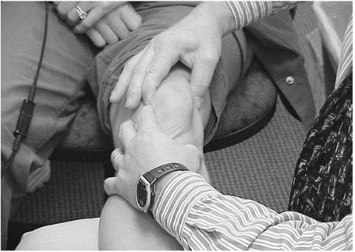
Apprehension test (Figure 11.2): in patients with spontaneous reduction or subluxation, patellar stability is assessed by placing the patient supine with the knee flexed over the examiner’s thigh (approximately 10°). The examiner applies pressure to the medial patella forcing it laterally; pain and increased motion are suggestive of patellar instability.
Crs:
Dislocation requires reduction.
Following reduction, pain at medial retinaculum and lateral patella subside over 4-8 w.
Complc:
Stay updated, free articles. Join our Telegram channel

Full access? Get Clinical Tree


