Abstract
Introduction
Pain is the main problem in patients suffering from cerebral palsy, particularly in adults. The upper limbs are affected in 25% of cases. Here, we report the case of a patient with Kienböck’s disease.
Method
Clinical case and literature review. A 28-year-old man suffering from dystonic quadriplegia consulted for progressively worsening pain in the right wrist. Kienböck’s disease was diagnosed and conservative treatment with botulinum toxin in the flexor carpi radialis recommended. A good result was obtained with a decrease in pain. This result was still present two years later.
Discussion
Although few references are made to it in literature, Kienböck’s disease in cerebral palsy is probably underestimated. Maintenance of the wrist in a permanent flexed position and muscular hypertonia may be risk factors. Knowledge of this particular clinical picture will enable it to be detected promptly and thus enable conservative treatment to be organised with a maximum chance of therapeutic success, preventing the need for surgery.
Résumé
Introduction
La douleur est le principal motif de consultation des personnes atteintes de paralysie cérébrale, notamment dans la population adulte. L’atteinte des membres supérieurs est retrouvée dans un quart des cas. Nous rapportons ici le cas d’un patient souffrant d’une ostéonécrose du lunatum connue sous le nom de maladie de Kienböck.
Méthode
Présentation d’un cas clinique et revue de la littérature.
Observation
Un patient de 28 ans présentant une tétraparésie dystonique consulte pour une douleur de poignet de rythme mécanique et d’aggravation progressive. Après le diagnostic de maladie de Kienböck de stade 2 de Lichtman, un traitement conservateur par immobilisation avec traitement associé de la spasticité du fléchisseur radial du carpe par toxine botulique permet d’obtenir une nette amélioration, avec diminution de la douleur de 80 % selon le patient. Le bénéfice thérapeutique persiste à deux ans, avec des injections itératives de toxine botulique. Sur le plan radiologique, aucune modification n’est observée à deux ans.
Discussion
La maladie de Kienböck chez les personnes atteintes de paralysie cérébrale est peu décrite dans la littérature mais probablement sous-estimée. L’attitude permanente en flexion du poignet et l’hypertonie musculaire pourrait être des facteurs de risques. La connaissance de ce tableau clinique particulier doit permettre de l’évoquer précocement et de mettre ainsi en place un traitement conservateur avec un maximum de chance de succès thérapeutique et ainsi d’éviter un geste chirurgical.
1
English version
Pain is the main health problem reported by adult sufferers of cerebral palsy. Localisation in the upper limbs is reported in 25% of cases, with a frequent context of overuse . Kienböck’s disease is an ischaemic necrosis of the lunate bone, posing the same etiological problem as other idiopathic necroses: none of the many explanations set forth in literature has yet provided definite proof of its value . Among these, hyperflexion of the wrist resulting from spasticity of the flexors was detected in sufferers of neurological ailments . Spasticity and dystonia are constants in cerebral palsy, the current term for cerebral motor disability; it could therefore be assumed that Kienböck’s disease is frequently found in this context. Even if two series report rates of 2.7% and 10% in patients presenting with cerebral palsy , the number of cases described in literature remains extremely slight. Here, we will report on an observation of a young cerebral palsy sufferer. We will then compile another summary of the information provided in literature from this case.
1.1
Observation
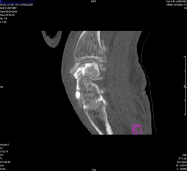
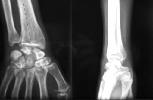
M.D., a male of 28 years suffering from dystonic quadriplegia in the context of cerebral palsy, with bilateral flexion of the wrists from a clinical viewpoint in particular. He consulted with a progressive pain in his right wrist, with tumefaction of the carpus. M.D. is right-handed, entirely autonomous, with no previous asymmetry noted on the articular amplitudes of the wrists with a 40° bilateral extension limit. The pain developed after 15th May 2006, was strictly unilateral and became permanent leading to an X-ray in September 2006. This led to the diagnosis of Lichtman stage 2 osteonecrosis of the lunate bone, which would be confirmed by CT scan with a fragmentary aspect of the lunate bone ( Figs. 1 and 2 ). Negative ulnar variance was also noted ( Fig. 1 ) in the standard radiological assessment. A surgical procedure to shorten the radius was recommended on the therapeutic level due to the inequality of the length of the two bones in the forearm. However, fear of increasing the handicap led to conservative treatment being proposed in the first instance. An injection of botulinum toxin into the flexor carpi radialis was proposed in conjunction with immobilisation of the wrist using a splint. Toxin A was injected on 13th December: 100 U Botox in 1 ml via tracking by stimulation in the flexor carpi radialis. Distinct improvement was noted after 15 days, with a distinct decrease in pain which only persisted when extended, but M.D. no longer took analgesics. He did not complain of loss of strength nor loss of function. The injections were repeated every three months with persistence of the benefit; the splint was no longer worn continuously. After two years, M.D. no longer took analgesics. He estimated that there had been a 70% improvement and enjoyed normal functioning of the right wrist again. He wore his splint during his packaging job at a sheltered workshop. An X-ray showed images of osteonecrosis without lunate bone collapse.

1.2
Discussion
Kienböck’s disease is a rare pathology which most frequently affects young men of between 20 and 40 years of age.
Pain is the main reason for consultation in sufferers of cerebral palsy . The vertebral column and lower limbs are the most frequent locations. It is reported that the upper limbs are affected in 25% of patients . The most frequently encountered problems are articular overwork syndromes, in particular in conjunction with the usage of walking sticks or wheelchairs, and pain related to spasticity and dystonia. Few studies have been devoted to the incidence of Kienböck’s disease in cerebral palsy. This is 2.7% according to Joji et al. . He actually found six cases in a systematic radiological study involving 110 patients suffering from cerebral palsy, with one case of bilateral affliction. Only four patients presented symptoms, one of whom was the patient presenting with a bilateral affliction who only suffered unilaterally. Both wrists had previously been painful but were asymptomatic at the time of the study. Rooker and Goodfellow found an incidence of 10% in a population of 50 patients. Of the 12 patients presenting with wrist pains, five actually presented with Kienböck’s disease . These two series are the only ones in literature within the context of cerebral palsy; the other cases described are isolated cases as our experience shows us. In a radiological study, Nishioka et al. did not find a single case in 96 patients. It should be noted that he found an 18.2% negative ulnar variance rate.
All the observations described relate to patients who tend to be adults, presenting with a spastic and athetosic affliction with palmar flexion of the wrist. The affliction is not linked to the dominant hand and a short ulna was most frequently observed in standard radiography, as in our observation.
Osteonecrosis of the lunate bone is apparent from the occurrence of a mechanically rhythmic pain which progressively worsens, with subsequent appearance of stiffness. Radiological assessments are normal in the first instance, but subsequently develop into condensation of the bone and fragmentation. In the first instance, scintigraphy enables affirmation of the location on the lunate bone, but although its sensitivity is good, its specificity is less so. MRI is more successful and shows an ischaemic aspect of the bone at an early stage, with loss of the T1 signal, focused on the supero-external part to begin with. Loss of the T2 signal is a symptom of severity. CT scan shows fragmentation of the bone which is not necessarily shown with standard radiography . Arthro-CT scan provides good visualisation of any possible osteoarthritis.
The etiology of Kienböck’s disease is multifactorial. The notion of trauma upon hyperextension of the wrist may be found in the patient’s case history, in the same way as the notion of repetitive microtrauma.
Extracarpal hyperpressure is often found in the course of the disease. This may be the result of a short ulna, an anomaly of the radial slope or an overhang of bone under the ulna. In spastics, permanent contraction of the flexors in the wrist is a factor which causes hyperpressure and decreased venous return due to the flexed position of the wrist which favours the occurrence of osteonecrosis. A simple chance association cannot be completely ruled out, of course.
This develops into a chronically painful picture with stiffening of the wrist and functional impotence. From a radiological viewpoint, destabilisation of the carpus appears with collapse of the lunate bone and localised or generalised arthrosic development.
On a therapeutic level, treatment may be medical but suggested therapy is most often surgical . Medical treatment consists of immobilisation of the wrist for analgesic purposes. Greene thus described a favourable development in a 10-year-old cerebral palsy sufferer, with complete immobilisation of the wrist at 10° extension for 15 weeks, followed by intermittent immobilisation in a splint for a further 15 days when a walking frame was used. Radiological assessment at the age of 17 years was normal, with disappearance of the painful symptomatology.
For many authors, surgical treatment yields a better result: levelling of the two joints in the forearm by shortening the radius or lengthening the ulna is recommended when the bones in the forearm are unequal in length at stage 2. Shortening of the capitum has also been recommended, as has replacement of the lunate. Arthrodesis of the carpus may also be performed as may resectioning of the proximal row of the carpus. These latter indications mainly relate to stages 3 and 4. For Leclercq and Xarchas , this procedure simultaneously permits improvement of the spasticity of the wrist flexors within the context of cerebral palsy.
The prompt commencement of medical treatment seems to be a vital element in therapeutic success . In spastic or dystonic patients, efficient immobilisation can only be achieved with treatment tailored to spasticity to break the wrist flexing pattern. Botulinum toxin is highly indicated here . Within the context of cerebral palsy, diagnosis must therefore be systematically made in the face of painful affliction of the wrist by performance of a standard X-ray, taking into account the absence of clinical specificity. If the radiological assessment is normal, a clinical and radiological monitoring programme must be implemented. The possible presence of a short ulna requires even greater vigilance. An MRI may be discussed in the first instance to obtain a definite diagnosis in the event of extremely invalidating pain and normal radiography.
All in all, Kienböck’s disease is probably underestimated in people affected by cerebral palsy. Knowledge of this particular clinical picture should enable it to be detected early, thus enabling a conservative treatment programme to be implemented where botulinum toxin plays an important part, with a maximum chance of therapeutic success.
2
Version française
La douleur est le principal problème de santé dont se plaignent les adultes atteints de paralysie cérébrale. La localisation aux membres supérieurs est rapportée dans 25 % des cas avec fréquemment un contexte de sur-utilisation . La maladie de Kienböck est une nécrose ischémique de l’os lunatum, posant le même problème étiologique que les autres nécroses idiopathiques : aucune des multiples explications proposées dans la littérature n’a encore fourni de preuve certaine de sa valeur . Parmi celles-ci, l’hyperflexion du poignet consécutive à la spasticité des fléchisseurs a été évoquée chez le sujet atteint d’affection neurologique . Spasticité et dystonie sont des constantes dans la paralysie cérébrale, dénomination actuelle de l’infirmité motrice cérébrale, on peut donc penser que dans ce contexte la maladie de Kienböck va fréquemment être retrouvée. Même si deux séries rapportent des taux de 2,7 et 10 % chez des patients présentant une paralysie cérébrale , le nombre de cas décrit dans la littérature reste extrêmement faible. Nous rapportons ici une observation chez un jeune patient atteint de paralysie cérébrale. À partir de ce dossier, nous referons une synthèse des données de la littérature.
2.1
Observation
M.D., homme de 28 ans, est atteint d’une quadriplégie dystonique dans le cadre d’une paralysie cérébrale, avec notamment cliniquement des poignets en flexion de façon bilatérale. Il consulte pour une douleur du poignet droit d’installation progressive avec tuméfaction du carpe. M.D. est droitier, entièrement autonome, auparavant aucune asymétrie sur les amplitudes articulaires des poignets n’était notée avec une limitation bilatérale d’extension à 40°. La douleur évolue depuis le 15 mai 2006, strictement unilatérale, et est devenue permanente amenant à la réalisation d’un cliché radiologique au mois de septembre 2006 conduisant au diagnostic d’ostéonécrose du lunatum stade 2 dans la classification de Lichtman qui va être confirmé par un scanner avec un aspect de fragmentation du semi-lunaire ( Fig. 1 et 2 ). Au bilan radiologique standard, une variance ulnaire négative est également notée ( Fig. 1 ). Au niveau thérapeutique, en raison de l’inégalité de longueur des deux os de l’avant-bras, un geste chirurgical de type raccourcissement du radius est évoqué. Néanmoins, la crainte d’une majoration du handicap amène à proposer un traitement conservateur dans un premier temps. Une injection de toxine botulique au niveau du fléchisseur radial du carpe est proposée associée à une attelle d’immobilisation du poignet. L’injection de toxine A est effectuée le 13 décembre : 100 U Botox dans 1 ml sous repérage par stimulation dans le fléchisseur radial du carpe. Après 15 jours, une nette amélioration est relevée, avec une nette diminution de la douleur qui ne persiste qu’en extension, mais M.D. ne prend plus d’antalgique. Il ne se plaint pas de perte de force, ni de perte fonctionnelle. Les injections sont renouvelées tous les trois mois avec persistance du bénéfice, l’attelle n’est plus portée en continu. À deux ans, M.D. ne prend plus d’antalgique, il s’estime amélioré à 70 % et a retrouvé une fonction normale du poignet droit, l’attelle est portée durant l’activité professionnelle de conditionnement au CAT. Le contrôle radiologique retrouve les images d’ostéonécrose sans affaissement du semi-lunaire.
Related posts:
 Do patients have any special medical or rehabilitation difficulties after a craniectomy for malignant cerebral infarction during their hospitalization in a physical medicine and rehabilitation department?
Claw toes in hemiplegic patients after stroke
The use of dry powder inhaler devices by elderly patients suffering from chronic obstructive pulmonary disease
Patients in a permanent vegetative state or minimally conscious state in the Maine-et-Loire county of France: A cross-sectional, descriptive study
The management of stroke patients. Conference of experts with a public hearing. Mulhouse (France), 22 October 2008
Evaluation of a wheelchair prototype with non-conventional, manual propulsion
Do patients have any special medical or rehabilitation difficulties after a craniectomy for malignant cerebral infarction during their hospitalization in a physical medicine and rehabilitation department?
Claw toes in hemiplegic patients after stroke
The use of dry powder inhaler devices by elderly patients suffering from chronic obstructive pulmonary disease
Patients in a permanent vegetative state or minimally conscious state in the Maine-et-Loire county of France: A cross-sectional, descriptive study
The management of stroke patients. Conference of experts with a public hearing. Mulhouse (France), 22 October 2008
Evaluation of a wheelchair prototype with non-conventional, manual propulsion
Stay updated, free articles. Join our Telegram channel

Full access? Get Clinical Tree


