Introduction
Nerve conduction studies are technical procedures used to objectively assess the functional state of the peripheral neuromuscular system. Standardized technical procedures increase the reliability of the studies. The standardized procedures presented in this book represent the consensus of experts who routinely perform nerve conduction studies and whose studies have been published in peer-reviewed journals.
Basic Nerve Conduction Studies: General Information
The study of nerve conduction assumes that when a nerve is stimulated electrically a reaction should occur somewhere along the nerve. The reaction of the nerve to stimulation can be monitored with appropriate recording electrodes. Direct recording can be done along sensory or mixed nerves. Indirect recording from a muscle can be used for motor conduction studies. Orthodromic and antidromic conduction can be studied, because stimulus propagation occurs both proximal and distal to the point of stimulation. Orthodromic conduction is the same direction as physiological conduction (e.g., sensory conduction toward the spinal cord and motor conduction away from the spinal cord). Antidromic conduction is propagation in the opposite direction. The time relationship between the stimulus and the response can be displayed, measured, and recorded.
Electrodes
Active (recording) and reference electrodes are used. The type of metal surface electrodes used is determined by the type of nerve response being studied.
Motor Response
Peripheral nerves may be stimulated by passing electrical currents through the skin, resulting in a synchronized muscle contraction. When recorded by surface electrodes, this is called the compound muscle action potential (CMAP). Motor responses are recorded over the muscle being studied. The active (recording) electrode (E1) should be placed over the motor point of the muscle so that a clear negative deflection (upward) is recorded when electrostimulation is applied to the nerve supplying that muscle. The reference electrode (E2) should be placed off the muscle on a nearby tendon or bone. Standard recording and reference electrodes are about 1.0 cm in diameter. The surface disc electrodes may be separate discs or fixed 2.0 to 3.0 cm apart in a plastic bar. For standardization or comparisons, E1 and E2 used in CMAP recording should be the same size.
Sensory Response
A compound nerve action potential produced by electrical stimulation of the afferent nerve may be recorded over peripheral sensory nerves in a number of areas. The reference electrode usually is the same type as the recording electrode, and in antidromic studies it is placed distal to the recording electrode. For the orthodromic recording, the reference electrode is placed proximal to the recording
electrode. Recording and reference electrodes used for sensory responses usually are surface electrodes with a spring ring, disc, or bar (Figs. 1-1 and 1-2). Disc, bar, clip, and ring electrodes may be used interchangeably for recording and stimulating.
electrode. Recording and reference electrodes used for sensory responses usually are surface electrodes with a spring ring, disc, or bar (Figs. 1-1 and 1-2). Disc, bar, clip, and ring electrodes may be used interchangeably for recording and stimulating.
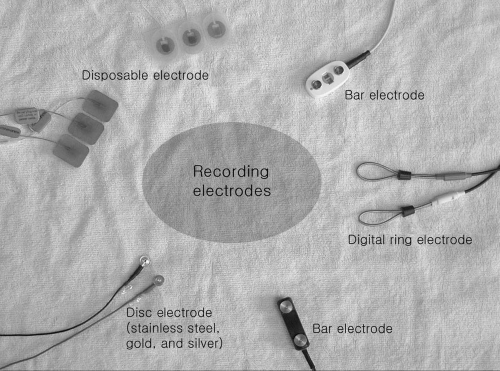 Figure 1-1. Different types of surface recording electrodes. |
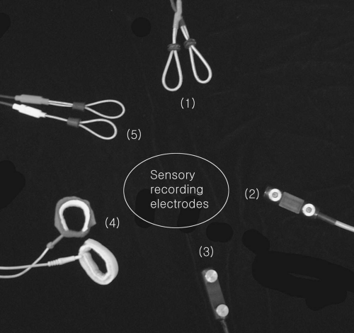 Figure 1-2. Sensory recording electrodes. |
Ground Electrodes
The ground electrode (Fig. 1-3) is a metal plate that provides a large surface area of contact with the patient. It serves as a reference zero potential and helps to reduce stimulus artifacts. The ground electrode usually is placed between the active electrode and the stimulator. It usually is larger than the recording and reference electrodes.
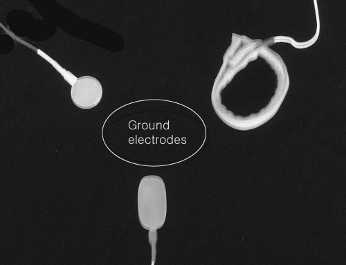 Figure 1-3. Ground electrodes. |
Stimulating Electrodes
Surface stimulation electrodes usually are two metal electrodes or felt pad electrodes placed 1.5 to 3.0 cm apart (Fig. 1-4). The use of this stimulator is standard
in conventional nerve conduction studies. However, the monopolar needle electrode also can be used for nerve stimulation. Its use has certain advantages: (a) a smaller stimulus intensity is required, (b) the nerve can be stimulated more selectively than with a surface stimulating electrode, and (c) nerves that lie anatomically deep can be stimulated (e.g., the spinal nerve roots or sciatic nerve in the sciatic notch).
in conventional nerve conduction studies. However, the monopolar needle electrode also can be used for nerve stimulation. Its use has certain advantages: (a) a smaller stimulus intensity is required, (b) the nerve can be stimulated more selectively than with a surface stimulating electrode, and (c) nerves that lie anatomically deep can be stimulated (e.g., the spinal nerve roots or sciatic nerve in the sciatic notch).
 Figure 1-4. Different types of stimulators. |
Techniques: General Considerations
A few general rules make nerve conduction velocity studies easy to perform and greatly reduce the number of examiner errors. Ensure that the following steps are taken:
Cleanse all recording, reference, ground, and stimulating electrodes after each use by washing with warm soapy water. Each electrode should then be dried completely.
Electrically test all electrodes for broken wires or defective contact points. If a defect is noted, repair or replace the electrode.
Ensure that the electrode site on the patient’s skin is clean and free of oil, grease, and soil. The site should be cleaned and abraded, as necessary, to reduce impedance at the electrode/skin interface.
Mark all recording and stimulating points clearly with visible ink.
Measure distances with a tape measure that is closely apposed to the skin and anatomic course of the nerve.
Position the cathode (negative pole) of the stimulating electrode toward the active (recording) electrode for most of the studies presented in this manual.
Ensure that the stimulus is adequate to evoke a motor or sensory response. In general, a stimulus is defined as any external agent, state, or change that is capable of influencing the activity of a cell, tissue, or organism. In clinical nerve conduction studies, an electric stimulus generally is applied to a nerve or muscle. The electric stimulus may be described in absolute terms or with respect to the evoked potential of the nerve or muscle. In absolute terms, the electric stimulus is defined by a duration (current pulse width) in milliseconds (ms), a waveform, and a strength or intensity measured in voltage or current (milliamperes). With respect to the evoked potential, the stimulus may be graded as subthreshold, threshold, submaximal, maximal, or
supramaximal. The threshold stimulus is that stimulus sufficient to produce a detectable response. Stimuli less than the threshold stimulus are termed subthreshold. The maximal stimulus is the stimulus intensity after which a further increase in the stimulus intensity causes no increase in the amplitude of the evoked potential. Stimuli of intensities below this level but above threshold are submaximal. Stimuli of intensities greater than the maximal stimulus are termed supramaximal. Ordinarily, supramaximal stimuli are used for nerve conduction studies. By convention, an electric stimulus of about 20% greater voltage/current than required for the maximal stimulus should be used for supramaximal stimulation. The frequency, the number, and the duration of a series of stimuli should be specified on the reporting form.
Latency time (ms) for motor responses is measured from the shock artifact to the initial negative deflection (upward) of the response from the isoelectric baseline of the video display apparatus (Fig. 1-5).
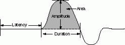
Figure 1-5. CMAP parameters.
With the motor nerve conduction velocity (NCV) technique, the reference and ground electrodesRelated posts:

Stay updated, free articles. Join our Telegram channel

Full access? Get Clinical Tree


