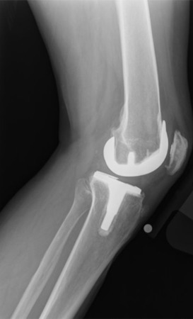
Instability is one of the most common causes of failure after total knee arthroplasty. Although there are several contributing causes, surgical error and poor implant design selection contribute. For this reason, an accurate diagnosis is fundamental and is largely based on a thorough history and physical examination. In general, tibiofemoral instability can be classified into 3 different patterns: flexion instability, genu recurvatum, and extension instability. In this article, these 3 patterns are reviewed in greater depth.
Key points
- •
Instability is not always associated with discomfort, and the diagnosis is largely based on a complete history and physical examination.
- •
In cruciate-retaining designs, flexion instability may be caused by surgical errors or posterior cruciate ligament failure. In posterior-stabilized designs, the cause is usually unbalanced flexion-extension gaps.
- •
Genu recurvatum is a rare complication caused by underlying conditions like quadriceps weakness, paralysis, bone deformities, previous high tibial osteotomies, and plantar foot flexion.
- •
Extension instability can produce rectangular or trapezoidal gaps and thus symmetric or asymmetrical instability, respectively.
Introduction
Instability is one of the most important causes of failure after total knee arthroplasty (TKA), accounting for 10% to 20% of all knee revision procedures. In the United States and Australia, it is one of the most common causes of late revisions. It has also been reported as one of the most common early complications, found to be as high as 26% in the first 5 years after surgery, and the second most common cause of revision after infection.
Instability is not always associated with discomfort. In addition, the causes are various and include surgical error and poor design selection. The treatment of instability is based on an accurate diagnosis, founded on a thorough history and physical examination. Onset of signs and symptoms must also be investigated carefully. For instance, a cruciate-retaining (CR) TKA can be unstable in flexion after a period of wellness secondary to a late posterior cruciate ligament (PCL) rupture.
In general, tibiofemoral instability can be classified into 3 different patterns: flexion instability, genu recurvatum, and extension instability.
Evaluation
The first step is a thorough history and physical examination, focusing on the following aspects :
- •
Original indication for surgery
- •
Previous deformity or contracture
- •
Previous knee surgeries
- •
Wound complications
- •
Current sense of instability
- •
Recurrent effusions
- •
Time of Onset
- •
Pain localization
The examination should be accurate and focused not only on the knee but also on extra-articular causes of instability. Before addressing the knee, it is useful to search for global or local neuromuscular disorders, hip or ankle deformities, and areas of tenderness (i.e., pes anserine and Gerdy’s tubercle).
Varus-valgus testing of the knee should be performed in full extension and at 30° and 90° of flexion to evaluate additional knee stabilizers. Anteroposterior (AP) laxity should be evaluated in both directions with anterior and posterior drawer tests. A useful test is performed with the patient sitting on the examination table, with the knee flexed at 90° and the foot dangling to assess the extent to which the flexion gap increases.
When evaluating a painful TKA, the surgeon must first exclude infection as a primary cause of the failure. A contemporary approach has been proposed by the Musculoskeletal Infection Society and is supported by other studies. Diagnostic tests include erythrocyte sedimentation rate, C-reactive protein, and an arthrocentesis looking at synovial white blood cell count, differential, and cultures. For this reason, an arthrocentesis is always performed to evaluate the articular fluid and rule out infection. Instability can present with hemarthrosis secondary to intra-articular microtrauma and is another diagnostic clue.
Imaging analysis
Radiographic analysis of the knee should investigate implant positioning, limb alignment, and component position. Obligatory radiographs include AP, lateral, full-length weight-bearing, and patellar views. AP radiographs can be completed with varus-valgus stress images to assess the status of the collateral ligaments and if deformities are reducible.
Lateral views can be performed in full extension, 90° of flexion, and full flexion. In the different views, it is possible to measure the tibial translation on the femur, implant positioning, flexion gap, and tibial slope. Full-length radiographs are useful in assessing component positioning compared with anatomic and mechanical axes of the femur and tibia. Radiographs should always be compared with the preoperative and immediate postoperative radiographs to understand the evolution of disease.
Computed tomography scans may be considered when malrotation of the components is suspected. Although rarely indicated, MRI can be used for soft tissue evaluation and component rotation. However, this study is technically demanding and difficult to obtain even with metal artifact suppression technology.
Flexion instability
Flexion instability can be seen in patients without radiographic evidence of malalignment or loosening. This problem has been historically underdiagnosed, especially in patients with a CR implant. Flexion instability can also occur in patients with posterior-stabilized (PS) prostheses. The signs and symptoms may vary from simple discomfort to a complete knee dislocation.
Factors leading to instability in CR prostheses may include surgical error or late PCL failure. Surgical errors include a loose flexion gap (eg, undersized femoral component or excessive tibial slope) or a misdiagnosed previous PCL rupture. An excessively tight flexion gap has to be avoided as well, because the patient may be more prone to stiffness postoperatively. A subsequent manipulation and PCL overstretching when in flexion may then lead to late PCL rupture and flexion instability. In chronic cases, the PCL can deteriorate, leading to flexion instability. Either way, posterior sag of the tibia is clinically observed against gravity and at 90° of flexion.
In PS implants, frank knee dislocations are prevented by the polyethylene post and the femoral cam mechanism. However, an unbalanced flexion gap can affect stability, leading to anterior tibial translation and instability. In 2005, Schwab and colleagues analyzed flexion instability without frank dislocation in 10 patients with a PS design and reported the following classic symptoms:
- •
Sense of instability without giving away
- •
Difficulty in ascending and descending stairs
- •
Recurrent knee swelling
- •
Anterior knee pain and tenderness
During clinical examination, anterior tibial translation occurs with the knee flexed at 90°. In addition, multiple areas of soft tissue tenderness (i.e., pes anserine, peripatellar region, and hamstrings tendons) and recurrent knee swelling (i.e., hemarthrosis) are appreciated.
In 2014, Abdel and colleagues correlated the radiographic findings of 60 patients revised for isolated flexion instability. These investigators found a strong correlation with decreased condylar offset by 4 mm ( P <.001), distalization of joint line by 6 mm ( P <.001), and increased tibial slope up to 5° ( P <.001) ( Fig. 1 ) to be predictive of flexion instability.


Stay updated, free articles. Join our Telegram channel

Full access? Get Clinical Tree


