Narrowing of the femoral neck after metal-on-metal hip resurfacing arthoplasty has been reported as a common radiologic feature, although its significance is still unknown. This study reports the presence and significance of neck narrowing in the first 500 consecutive Conserve® Plus metal-on-metal hip resurfacings in 431 patients.
Metal-on-metal hip resurfacing is an attractive option for young patients, as it leaves more femoral bone stock if revision surgery is necessary. It also restores normal anatomy and biomechanics of the hip, while providing near-normal proximal femoral anatomy and loading. Narrowing or thinning of the femoral neck after metal-on-metal hip resurfacing arthroplasty has been reported as a common radiologic feature. Although the long-term effects of neck narrowing are unknown, it has been speculated that neck narrowing does not result in adverse clinical or radiologic outcomes.
This study reports the incidence and significance of neck narrowing in the first 500 consecutive hips in the senior surgeon’s (H.C.A.) series with a relatively long follow-up and determines the cause where possible. The radiological features of neck narrowing were examined over time, as well as the clinical features and histology of retrievals of those hips that failed with neck narrowing.
Materials and methods
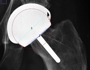
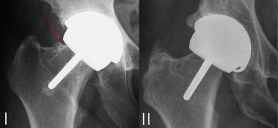
Between 1996 and 2002, the senior author (H.C.A.) implanted the first 500 consecutive Conserve® Plus metal-on-metal hip resurfacings (Wright Medical Technology, Inc, Arlington, TN, USA) in 431 patients (319 men, 112 women). The mean age at the time of surgery was 48.6 years, and the average follow-up was 95.9 (range, 1.4–161) months. Two patients were lost to follow-up. Neck narrowing was measured using a modified protocol used by Hing and colleagues. Immediate postoperative radiographs were compared with the most recent postoperative radiographs for narrowing of the femoral neck at the component-neck junction ( Fig. 1 ). All radiographs used were standardized anteroposterior (AP) views. Nonsuitable radiographs were excluded from the measurement (eg, femoral orientation not consistent with the rest of the series). Radiographic measurements of acetabular abduction and anteversion were made using the EBRA program (Einzel-Bild-Roentgen-Analysis, University of Innsbruck, Austria), and this program was also used to outline the femoral ball to find the center of the femoral head. Using the outline and the center of the femoral head, the Image J software (National Institute of Health, version 1.41) was used to measure the femoral ball diameter and the diameter of the neck at the component-neck junction. This procedure was performed by 2 independent observers (J.Y., K.M.T.). Radiographs that showed narrowing were evaluated at different time points over the series of follow-up visits to track changes in narrowing. The neck narrowing measurements were plotted against time to examine the progression of narrowing over the follow-up period. The femoral head diameter was used to calibrate the radiographs to measure the amount of neck narrowing in millimeters and to calculate the percentage of narrowing. Cases exceeding 10% narrowing were further classified depending on whether the narrowing involved most of the neck or was caused by the presence of a more localized triangular area or “bite,” with loss of bone at the component-neck junction and sclerotic line at the bite ( Fig. 2 , Table 1 ). Furthermore, the narrowing was assessed as either symmetric (both inferior and superior narrowing) or eccentric (mostly inferior or mostly superior narrowing). This assessment was done by the senior author (H.C.A.).


| Category | Description |
|---|---|
| I | Neck narrowing with a bite at the component-neck junction |
| II | Neck narrowing involving most of the neck |
After the initial analysis of neck narrowing, we performed a secondary biomechanical analysis similar to that of Joseph and colleagues to measure the abductor moment arm (AMA) and body moment arm (BMA) in 301 preoperative and 478 postoperative radiographs to calculate the hip ratio (HR), which indicates the force exerted by the abductor muscles and body weight to maintain equilibrium in the 1-leg stance ( Fig. 3 ). In addition, the stem shaft angle (SSA) was calculated for each hip. Radiographs without the entire AP view of the pelvis or with incomplete view of the greater trochanter were excluded from the biomechanical analysis. For the preoperative biomechanical analysis, 279 radiographs were available for the non–neck narrowing group and 22 for the neck narrowing group. For the postoperative biomechanical analysis, 446 radiographs were available for the non–neck narrowing group and 25 were available for the neck narrowing group.
The difference in preoperative and postoperative HR was calculated to assess changes in hip biomechanics. Clinical and radiologic variables including the underlying diagnosis, biomechanical and radiographic parameters (neck narrowing, abduction, anteversion, contact-patch-to-rim [CPR] distance as described by Langton and colleagues, SSA, preoperative and postoperative HR, change in HR, postoperative AMA, postoperative BMA), demographic data (age, height, weight, body mass index [BMI], femoral head size), and patient assessment scores (Harris hip score [HHS], University of California Los Angeles [UCLA] activity score) were compared between the neck narrowing and non–neck narrowing groups.
Two-tailed Student t -tests were used to compare parametric data. Mann-Whitney-U tests were used to compare nonparametric data. A P value of less than 0.05 was deemed to be significant. χ 2 Analysis with Fisher exact test correction was used to compare the ratio of failure with nonfailure cases between the groups with and without neck narrowing.
Retrieved implants were examined at the Implant Retrieval Laboratory at the Los Angeles Orthopaedic Hospital. Upon retrieval, the specimens were fixed in formalin. The retrieved implants were subsequently cleaned, photographed, and examined grossly. Then the femoral component and associated femoral neck were sectioned using an EXAKT saw (EXAKT Advanced Technologies, Norderstedt, Germany) into 2.5-mm slices, which were then radiographed. These slices were removed from the metal, decalcified, and processed into paraffin for routine histologic examination and staining by hematoxylin-eosin. The cut sections that included the femoral neck, neck implant junction, and resurfaced bone portions were examined for osteoclastic and osteoblastic activity, as well as viability of the bone, which was assessed by the presence of nuclei in the bone. Soft tissues adhering to the neck and interfacial membranes were examined for the presence of inflammatory cells and wear debris.
Results
Twenty-five hips in 22 patients were identified in the first 500 hips to have neck narrowing greater than 10%, which gives an incidence of 5%. In hips with neck narrowing, there were 11 males (13 hips, 52%) and 11 females (12 hips, 48%). In hips without neck narrowing, there were 311 males (358 hips, 77.2%) and 106 females (117 hips, 25.2%). There was a significant difference in gender between the two groups (Chi-squared, P = .001), with a higher proportion of females in the neck narrowing group, occurring in 3.6% of male hips and 10.3% female hips.
There was no difference in the underlying diagnosis among cases with neck narrowing compared with the rest of the cohort (χ 2 , P = .36) ( Table 2 ). There was 20.1% (10.8%–38.7%) narrowing in the neck narrowing group, and the narrowing measured 7.1 mm (3.5–14.2 mm) at the last follow-up (mean, 101 months).
| Etiology | No Neck Narrowing | Neck Narrowing | Total |
|---|---|---|---|
| OA | 300 (63.2%) | 15 (60%) | 315 (63%) |
| ON | 39 (8.2%) | 2 (8%) | 41 (8.2%) |
| DDH | 55 (11.6%) | 2 (8%) | 57 (11.4%) |
| Posttrauma | 38 (8%) | 1 (4%) | 39 (7.8%) |
| Inflammatory | |||
| Inflammatory OA | 9 (1.9%) | — | 9 (1.8%) |
| Ankylosing Spondylitis | 3 (0.6%) | 1 (4.0%) | 4 (0.8%) |
| JRA | 3 (0.6%) | — | 3 (0.6%) |
| RA | 4 (0.8%) | — | 4 (0.8%) |
| RD | 1 (0.2%) | — | 1 (0.2%) |
| Childhood disorders | |||
| LCP | 12 (2.5%) | 1 (4%) | 13 (2.6%) |
| SCFE | 7 (1.5%) | 2 (8%) | 9 (1.8%) |
| Other | |||
| Melorheostosis | 1 (0.2%) | — | 1 (0.2%) |
| Epiphyseal Dysplasia | 3 (0.6%) | 1 (4%) | 4 (0.8%) |
| Total | 475 | 25 | 500 |
There were no differences in the age, weight, BMI, SSA, HHS, or UCLA activity score ( Table 3 ). Height and femoral component size were significantly smaller in the neck narrowing group compared with the non–neck narrowing group only in men ( P = .0001 and P = .02, respectively).
| No Neck Narrowing (n = 475) | Neck Narrowing (n = 25) | P Value | |
|---|---|---|---|
| Age | 49 (15.3–78.1) | 46 (18.2–68.1) | .19 |
| Height (cm) (male) | 178.8 (158–198) | 171 (157–183) | .0001 |
| Height (cm) (female) | 164.4 (140–183) | 164.3 (155–178) | .92 |
| Weight (kg) (male) | 89.1 (57–164) | 82.5 (57–99) | .24 |
| Weight (kg) (female) | 67.8 (42–107) | 66.3 (50–103) | .70 |
| BMI (kg/m 2 ) (male) | 27.8 (18.4–46.4) | 28.1 (22.3–33.4) | .51 |
| BMI (kg/m 2 ) (female) | 25.0 (17.5–42.3) | 24.5 (19.0–32.5) | .88 |
| Female Femoral Head Size (mm) | 42 | 42 | .61 |
| Male Femoral Head Size (mm) | 48 | 46 | .02 |
| HHS | 93 (40.9–100) | 89 (52.9–99.9) | .08 |
| UCLA activity | 7.3 (3–10) | 7.2 (4–10) | .51 |
There were no significant differences in acetabular abduction, anteversion, or SSA ( Table 4 ). The neck narrowing group had a significantly lower CPR ( P = .04) and postoperative BMA ( P = .02). There were no significant differences in the postoperative AMA and HR.
| Non Neck Narrowing | Neck Narrowing | P Value | |
|---|---|---|---|
| Abduction | 43.4° (22°–71.5°) | 45° (16.2°–65.7°) | .39 |
| Anteversion | 16.9° (2.4°–51.2°) | 17.7° (3.0°–39.6°) | .98 |
| CPR (mm) | 14.7 (0.9–23.8) | 13.8 (3.2–21.3) | .04 |
| SSA | 137° (110°–163°) | 136° (125°–147°) | .59 |
| Preoperative HR a | 0.54 (0.31–0.99) | 0.55 (0.40–0.68) | .91 |
| Postoperative AMA (mm) b | 51 (32–70) | 49 (35–60) | .77 |
| Postoperative BMA (mm) b | 87 (64–102) | 84 (72–91) | .02 |
| Postoperative HR b | 0.59 (0.35–0.79) | 0.59 (0.46–0.73) | .20 |
| Change in HR a | −0.05 (−0.56–0.45) | −0.04 (−0.25 to 0.12) | .64 |
a For preoperative biomechanical analysis, 279 radiographs were available for the non–neck narrowing group and 22 were available for the neck narrowing group.
b For postoperative biomechanical analysis, 446 radiographs were available for the non–neck narrowing group and all 25 were available for the neck narrowing group.
Six hips were classified into the first category, with the bite occurring at the component-neck junction ( Fig. 4 , Table 5 ). Case 1 was an 18-year-old patient with severe femoral cystic degeneration secondary to Legg-Calvé Perthes disease; the component was seated on the superior neck and implanted with a cemented stem. The hip recently loosened on the acetabular side after 10 years and is now pending revision ( Fig. 5 ). Case 4 had an area of osteopenia superiorly and developed a bite at the component-neck junction, which did not progress during the past 4 years. The patient is active and plays racquetball (UCLA activity score 10) ( Fig. 6 ). Case 5 had severe bone stock deficiency with large defects and osteopenia inferiorly. The bite also occurred inferiorly; however, the narrowing is mostly taking place superiorly. Although narrowing is progressive, the patient is doing well at 10 years ( Fig. 7 ).
| Symmetric | Inferior | Superior | Total | |
|---|---|---|---|---|
| Category I | 1 (4.2%) | 2 (8.3%) | 3 (12.5%) | 6 (25%) |
| Category II | 14 (58.3%) | 1 (4.2%) | 3 (12.5%) | 18 (75%) |
| Total | 15 (62.5%) | 3 (12.5%) | 6 (25%) | 24 a |
a One case of neck narrowing could not be determined because of excessive abduction and anteversion.
Eighteen hips were classified into the second category, in which the narrowing involved most of the neck, not just an acute narrowing close to the component-neck junction ( Fig. 8 ). Although most have stabilized, there is 1 case that continued to narrow until it failed from femoral loosening (case 11) and 1 that continues to narrow (case 21) and may require revision for fluid-filled adverse local tissue reaction (ALTR) ( Fig. 9 ). Cases 22 and 23 (a 40-year-old male patient) had bilateral osteonecrosis and failed coring and considerable femoral bone loss at surgery. The necks narrowed progressively but stabilized by 5 years, with no further progression. The right hip demonstrates condensation of the medial cortical bone and is doing well at 12.5 years postoperation (UCLA pain, walking, and function scores of 10, activity score of 6).
All 7 failures in the neck narrowing group occurred in category II, and revision has been recommended for 2 hips with fluid-filled ALTRs. The 7 revised hips in the neck narrowing group were in category II, 5 of which were symmetric, 1 of which was narrowed inferiorly, and 1 of which was not able to be determined as a result of the cup obstructing the inferior view of the component-neck junction. There were 2 revisions for acetabular loosening (after an average of 105.9 months), 1 for femoral neck fracture after 50 months, 1 for a fluid-filled ALTR and high metal ions after 120 months, and 3 for femoral loosening (after an average of 84.4 months). One woman had bilateral femoral neck narrowing, and both sides failed because of femoral loosening. Both sides had extensive femoral cystic degeneration preoperatively and were performed with first-generation technique using a small femoral size (both 38 mm); these hips were loose radiographically (lucent lines around the stem) before the neck narrowed symmetrically. The component then loosened further and tipped into the varus.
In hips without neck narrowing, there were 34 revisions: 1 for acetabular loosening, 1 for recurrent dislocation, 20 for femoral loosening, 5 for neck fractures, 1 for sepsis, 4 for osteolysis, 1 for component size mismatch, and 1 for socket loosening with intraoperative pelvic central fracture with unstable initial fixation. The incidence of failure was significantly higher in the neck narrowing group (Fisher exact, P = .003), and the difference in survivorship between the groups were significant (log-rank test, P = .001) ( Fig. 10 ). The neck narrowing group had a lower survivorship rate of 86.7% at 7.7 years compared with 93.6% in the non–neck narrowing group.


