Humeral Shaft Fracture Stabilization with Elastic Nails
Nathan W. Skelley
J. Eric Gordon
DEFINITION
Humeral shaft fractures comprise approximately 2.5% of all traumatic fractures in children.11
Nearly all humeral diaphyseal fractures in children can commonly be treated nonoperatively with bracing and sling support.9, 10
Titanium elastic nails can be used to stabilize selected humeral shaft fractures in children and adolescents from the distal metaphysis to the proximal humeral physis.
ANATOMY
Proximally, the neurovascular bundles are located near the axilla, however, distally, the ulnar nerve and radial nerve pass in proximity to the medial epicondyle and lateral condyle, respectively (FIG 1).
If antegrade nails are to be used, the course of the axillary nerve is important as it passes lateral to the proximal humerus from posterior to anterior approximately 3.5 cm distal to the greater tuberosity in adults.5
PATHOGENESIS
The humerus is initially made of woven bone that is gradually replaced by stronger lamellar bone during childhood.
This developmental transition makes the bone prone to fracture with direct impact and falls on outstretched arms.
An increasingly active pediatric population and the increased prevalence of motorized sports has led to an increase in humeral shaft fractures.1, 3, 6, 11
Clinical examination and radiographs can assist in characterizing the injury mechanism.
NATURAL HISTORY
Most humeral shaft fractures are amenable to nonoperative treatment.
In young children, moderate shortening (<3 cm) is well tolerated as is angulation up to 30 degrees for fractures in the proximal third, 25 degrees in the middle third, and 20 degrees in the distal third.3, 6
Fractures in the distal one-fourth humeral metaphysis are more sensitive to angulation and should be maintained within 10 degrees of anatomic alignment.
PATIENT HISTORY AND PHYSICAL FINDINGS
Most humeral shaft fractures are identified by the common complaints of pain, discomfort, or disuse. In pediatrics, the patients may not clearly verbalize the pain. Therefore, discomfort or disuse of the extremity should encourage further evaluation.
High-energy trauma is commonly associated with shortening and angulation of the upper extremity. Soft tissue injuries are also common findings (FIG 2).
Humeral shaft fractures are commonly the result of mild trauma in the presence of pathologic lesions such as unicameral bone cysts.8
A complete distal neurologic and vascular examinations should be performed and documented to verify the patient is neurovascularly intact. In younger children, a careful motor examination is essential, as the sensory examination can be unreliable.
Examination with careful palpation of the remainder of the extremity should be performed because ipsilateral forearm, wrist, or shoulder injuries are not unusual (see FIG 2).

FIG 1 • Anatomy of the humerus with important neurovascular structures shown.
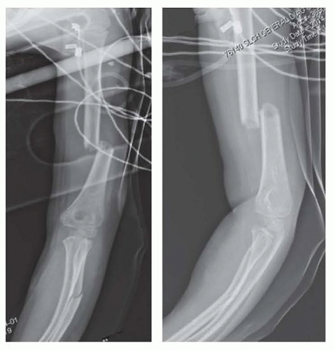
FIG 2 • Anteroposterior and lateral radiographs of the humerus in a 6-year-old girl demonstrating a displaced diaphyseal fracture sustained in a rollover motor vehicle accident. The patient also sustained an ipsilateral radius fracture, multiple head injuries, and ipsilateral tibia fracture.
Plain radiography is usually adequate to make a definitive diagnosis, although magnetic resonance imaging (MRI) can be occasionally helpful in distinguishing pathologic fractures through benign and malignant lesions.
IMAGING AND OTHER DIAGNOSTIC STUDIES
Two views (internally and externally rotated) of the humerus are usually sufficient to diagnose a humeral shaft fracture.
The joint above and below should be clearly visualized.
Concern on physical examination for injuries to the shoulder, forearm, or wrist should be evaluated by appropriate radiographs.
DIFFERENTIAL DIAGNOSIS
Pathologic fracture
Benign or malignant tumor
NONOPERATIVE MANAGEMENT
Humeral shaft fractures commonly do well functionally and cosmetically from nonoperative treatment.
Functional bracing, coaptation splints, hanging arm casts, or sling immobilization are common treatments.
Younger children have profound remodeling potential and can successfully remodel fractures with significant angulation up to 45 degrees.
SURGICAL MANAGEMENT
Surgical stabilization of humeral shaft fractures is indicated in certain scenarios including the following:
Open fracture
Inability to maintain adequate alignment by closed mean
Ipsilateral forearm fractures (floating elbow)
Closed head injuries to simplify nursing care
Polytraumatized patients, particularly with lower extremity fractures to facilitate mobilization out of bed by allowing upper extremity weight bearing6
The main goal of elastic nailing in humeral shaft fractures is to create a biomechanical stable reduction with axial, angular, translational, and rotational stability.
This is typically accomplished with two nails inserted through the metaphysis using a three-point application about the diaphyseal fracture site.
The nails are contoured by hand or with a plate-bending press to place the apex of the nail at the fracture site.
Elastic nailing is ideal for transverse and minimally comminuted fractures. Oblique diaphyseal fractures and unstable comminuted fractures can also be treated with flexible nailing because mild to moderate shortening rarely causes clinical problems.
Plate fixation requires a large incision, extensive dissection, and often requires exposure of neurovascular structures.
Preoperative Planning
A decision should be made between antegrade and retrograde nailing techniques.
Most fractures are treated with retrograde nailing. This technique allows stabilization of distal, midshaft, and proximal fractures with excellent stability.
The primary indication for antegrade nailing is soft tissue injury about the elbow requiring secondary procedures to obtain coverage.
Fractures in the distal and middle thirds of the humerus are best stabilized using retrograde nails placed from the medial and lateral sides of the elbow. Fractures in the proximal third of the humerus can be stabilized using two nails placed from the lateral side of the elbow, obviating the risk of ulnar nerve injury. FIG 3 shows incision sites and nail starting points.Related posts:

Stay updated, free articles. Join our Telegram channel

Full access? Get Clinical Tree


