 Hindfoot Nailing for Subtalar and Ankle Joint Fusion (Plantar Approach)
This approach is used only for hindfoot fusions that are to be treated by nailing. This technique is usually used for salvage of badly affected joints of the hindfoot to restore anatomic alignment. The procedure is often accompanied by a fibular osteotomy.
The skin of the sole of the foot is highly specialized, tough, and resilient. It responds to abnormal stresses by hypertrophying in the keratinized layer. The heel skin is especially thick. The approach for hindfoot nailing involves small incisions on the plantar aspect of the foot, carefully planned and usually performed on people with end-stage diseases of the hindfoot and ankle. Here the skin may be atrophic, especially with patients who have ischemic or neuropathic conditions.
Position of the Patient
Place the patient supine on the operating table (see Fig. 1-1). Partially exsanguinate the foot, either by elevating for a few minutes or by applying a soft rubber bandage loosely to the foot and binding it firmly to the calf. Then inflate a thigh tourniquet.
Landmarks and Incision
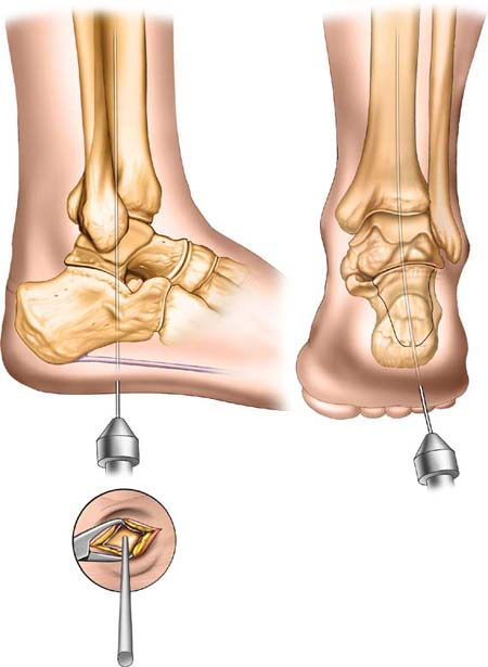
This approach is minimally invasive, and the small skin incision needs to be very accurately placed. Palpation of bony landmarks is insufficient and image intensification is usually used to help identify the internal bony architecture. To achieve a true lateral radiograph of the foot, roll the limb externally. To achieve a true anteroposterior radiograph of the ankle, bring the foot to the full dorsiflexed position (see Fig. 1-1).
Minimally invasive surgery demands careful preoperative planning, patient positioning, and very accurate skin incisions. Determine the position of the incision by using a lateral fluoroscopic image, an axial heel view, and an anteroposterior image. The starting point for the incision is determined by the intersection of two lines on the sole of the foot. The first line is drawn longitudinally through the ankle and hindfoot on the lateral image. This line runs through the center of the tibial medullary canal along its axis. The line crosses the talus and calcaneum to exit through the sole of the foot (Fig. 23-1A). The second line runs over the lateral column of the calcaneus and is determined on the longitudinal plantar view (Fig. 23-1B).
Stay updated, free articles. Join our Telegram channel

Full access? Get Clinical Tree


