General Complications of Arthroscopic Shoulder Surgery
Sumant G. Krishnan
Scott D. Pennington
Daniel E. Cooper
Wayne Z. Burkhead
INTRODUCTION
The evolution of both arthroscopic surgical techniques and equipment over the past 75 years has revolutionized shoulder surgery. Arthroscopy has evolved from a simple diagnostic tool to an invaluable instrument for minimally invasive shoulder reconstructions. The potentially devastating complications of open shoulder surgery, such as subscapularis repair failures and deltoid dehiscence, are minimized with arthroscopy but are not eliminated if attention to detail is lost (1). In truth, the overall complication rate of arthroscopic shoulder surgery is low but not insignificant.
A 1998 study from Madrid reported one surgeon’s arthroscopic complication rate of 10.63% (2). In 2002, Weber et al. (3) reported an overall complication rate in the published literature ranging from 5.8% to 9.5%. An exact rate is difficult to determine secondary to inaccuracies in reporting complications and disagreement over what constitutes a complication. For instance, some studies include postoperative shoulder stiffness as a complication, whereas others do not (2).
As Muller and Landsiedl stated, arthroscopy is not “band-aid surgery” (4). Specifically, problems can be encountered with patient positioning, surgical draping, and even arthroscopic portal placement. Patient selection, correct clinical diagnoses, and appropriate surgical indications remain critical to the success of any arthroscopic procedure, as some shoulder pathologies are not correctable by arthroscopic techniques alone. Anatomic structures are at risk and anatomic variants can cause confusion. Poor implant selection or placement may lead to intraoperative difficulty and possibly iatrogenic chondral damage. However, most complications of shoulder arthroscopy can be avoided by meticulous attention to detail.
PATIENT SELECTION
The appropriate patient selection for shoulder surgical procedures is covered comprehensively throughout this text, but several points with respect to shoulder arthroscopy bear mentioning. Although surgeons are attracted to the comprehensive visualization, scar cosmesis, and brisk return to function provided by shoulder arthroscopy, not all shoulder pathologies are amenable to arthroscopic procedures. Instability secondary to significant glenoid bony deficiency or large engaging Hill-Sachs lesions may require an open stabilization with an osseous transfer (iliac crest or Latarjet-Bristow procedure) (5). Massive and irreparable rotator cuff tears can be debrided or partially repaired arthroscopically, but large retracted and irreparable tears with significant fatty infiltration may require a tendon transfer. Failure to appreciate these clinical situations preoperatively will undoubtedly
lead to surgical frustration and potentially suboptimal patient outcomes.
lead to surgical frustration and potentially suboptimal patient outcomes.
ANESTHESIA CONCERNS
There have been several reports in the recent orthopaedic and anesthesia literature of anesthetic complications encountered during arthroscopic shoulder surgery (4). These include tracheal compression caused by periarticular fluid accumulation, visual loss and opthalmoplegia, pneumomediastinum from subcutaneous emphysema, spontaneous pneumothorax, and negative pressure pulmonary edema (6, 7, 8, 9, 10). These complications are exceedingly rare and hence difficult to screen for or prevent. The keys to managing (and possibly even preventing) these catastrophic situations are constant communication between the surgeon and anesthesiologist during the surgical procedure and a thorough knowledge of advance cardiac life support (ACLS) should the need arise for emergent perioperative resuscitation.
The main decision for patients and surgeons remains choice of anesthesia: general anesthesia versus regional anesthesia. Although the complications of general anesthesia are beyond the scope of this text, the most common complications of the interscalene brachial plexus block must be reviewed because this is the most widely used regional technique for arthroscopic shoulder surgery (3). Such complications most often involve palsy of the phrenic nerve (shortness of breath with hemidiaphragmatic paralysis), Horner’s syndrome (ipsilateral sympathetic blockade), and/or recurrent laryngeal nerve palsy (hoarseness and ipsilateral vocal cord paralysis). Although these complications are most often transient, they should be promptly evaluated when present by both the operating surgeon and the involved anesthesiologist.
Authors’ Preferred Anesthesia
We prefer to use general anesthesia for nearly every shoulder arthroscopy case. Although regional anesthesia is attractive, in our experience this technique is clearly dependent on the skill of the treating anesthesiologist and the incidence of transient vocal cord/laryngeal paralysis and/or hemidiaphragmatic paralysis is not negligible. At times, for postoperative pain control, we will combine a regional interscalene block with general anesthesia, although this may incur the risks inherent to both techniques.
PATIENT POSITIONING
Inappropriate patient positioning is perhaps the leading cause of complications both during and after arthroscopic shoulder surgery (11). Malpositioning can lead to significant upper and lower extremity neuropraxias as well as soft tissue injury away from the surgical site. The operating surgeon must be intimately involved during the installation of patients prior to shoulder arthroscopy, whether in the lateral decubitus position or the beach chair position. Surgeon participation at this level insures both the proper orientation of the patient for arthroscopic visualization as well as the optimal padding of bony prominences and susceptible neurovascular structures. Furthermore, preoperative planning by the operating surgeon may also dictate the type of positioning that may not be readily apparent to other physician or surgical assistants in the operating room. Arthroscopic posterior instability surgery may be more easily done in the lateral position, whereas the beach chair position may be preferred if the conversion to an open procedure is likely (3).
When placing the patient in the beach chair position, make certain the head is well-seated and secured in a fixed head holder with neutral position of the cervical spine (Fig. 11-1). Excessive pressure around the head can cause a neuropraxia of the posterior auricular nerve. The eyes should be free of any compression and the endotrachial tube should be directed away from the operative field. Flex the hips and knees comfortably such that the patient does not slide inferiorly during the procedure (Fig. 11-2). This also decreases both lumbosacral pressure and also the tension of the sciatic nerve. The neck must remain in a neutral position throughout the procedure to avoid any neuropraxias, and the anesthesiologist should be encouraged to frequently check the position of the head and neck during surgery (Fig. 11-3). Excessive flexion, extension, rotation,
or lateral bending of the cervical spine can lead to a brachial plexus injury (12,13).
or lateral bending of the cervical spine can lead to a brachial plexus injury (12,13).
 Figure 11-1 Beach chair position. |
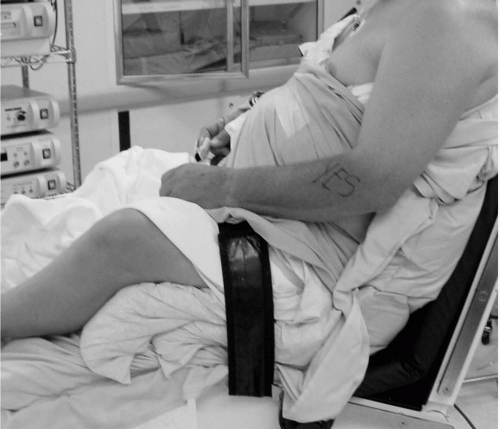 Figure 11-2 Appropriate flexion of hips and knees to relieve lumbosacral pressure and prevent traction on the long-tract nerves of the lower extremities. |
When installing the patient in the lateral decubitus position, the same parameters for the head and neck should be respected: neutral cervical spine position, avoidance of excessive pressure on the cranium or eyes, and flexion of both hips and knees (Fig. 11-4). Because in the lateral position the patient will be applying direct pressure to the contralateral half of the body, several other measures must be undertaken (3). An axillary roll should be placed under the contralateral upper ribs to prevent direct pressure through the axilla on the brachial plexus (though pressure-induced plexus palsy in the lateral position may still occur, even with appropriate axillary padding, if the patient has a cervical rib) (12,13). The contralateral peroneal nerve must be padded at the fibular head, as this is the nerve anatomically located in the most subcutaneous position that may be affected by prolonged direct pressure (14). The operative arm is often held in an abducted position by a longitudinal and perpendicular traction device. The appropriate amount of arm abduction to provide necessary visualization in the lateral position remains controversial and ranges from 0 to 90 degrees when combined with varying amounts of flexion. Published work has documented a reduced incidence of brachial plexus strain with increasing amounts of arm flexion (up to 90 degrees) and decreasing amounts of arm abduction (up to 0 degrees). We prefer to keep the arm in approximately 25 to 30 degrees of abduction in the scapular plane (approximately 30 degrees of forward flexion). Care must be taken to avoid excessive traction in either position (longitudinally or perpendicular to the glenohumeral joint). Neuropraxias have been documented by simple traction along the length of the operative arm. In addition, both abduction and traction changes the position of neurovascular structures at risk relative to the glenohumeral joint, and the potential for iatrogenic injury may increase if care is not taken when operating in the lateral position (14, 15, 16, 17, 18, 19, 20).
Related posts:
 Complications of Surgery to the Acromioclavicular and Sternoclavicular Joints
 Neurologic Injury
 Complications of Glenohumeral Arthrodesis
 Arthroscopy: Complications of Surgery for Instability
 Arthroscopy: Complications of Rotator Cuff Repair
 Complications of Arthroscopic Subacromial Decompression and Acromioclavicular Joint Resection
Stay updated, free articles. Join our Telegram channel

Full access? Get Clinical Tree


