Ganglia of the Hand
Christina F. Endress
Jonas L. Matzon
David R. Steinberg
CLINICAL PRESENTATION
Masses in the hand originate from either bone or soft tissues. Evaluation of a patient with a mass begins, as always, with a detailed history. History of the mass should include the rate of growth, changes in consistency or color, associated pain or neurologic symptoms, and any prior trauma to the area. The primary care physician should suspect malignant tumor if patients report rapid growth, night pain, and/or increase in pain. However, these symptoms can also occur with benign lesions.
While ganglia of the wrist are more common, the primary care physician may also encounter ganglia of the hand. Patients typically present with a smooth, firm mass that is sometimes tender and painful. The mass may fluctuate in size due to a one-way valve mechanism.1 Ganglia can spontaneously resolve; however, the recurrence rate is high. A higher recurrence rate has been observed in patients with a longer history and larger ganglia and when patients were operated by less experienced surgeons. Recurrence rates do not correlate with the ganglion location or the patient’s age or gender.2
Ganglia consist of thick, mucinous fluid composed of glucosamine, hyaluronic acid, albumin, and globulin that are surrounded by an acellular fibrous wall. The pathogenesis of ganglia is controversial. Some physicians believe that ganglia result from herniation of the synovial lining and collection of joint fluid. Others speculate that ganglia arise from synovial fluid leakage and subsequent irritation of the surrounding soft tissue and formation of a capsule. A few authors believe that ganglia may originate from mucoid degeneration of connective tissue and subsequent cyst formation.3 Hand ganglia typically manifest in one of three forms: flexor sheath cyst, proximal interphalangeal (PIP) mass, or mucous cyst.
The flexor sheath ganglion (volar retinacular or “seed” ganglion) usually arises on the volar surface of the hand between the A1 and A2 pulleys of the flexor tendon sheath (Fig. 49-1A). Most patients are young adults who complain of pain when grasping objects or making a tight fist.
Ganglia of the PIP joint usually affect middle-aged adults and may arise either from the extensor mechanism or directly from the PIP joint capsule (Fig. 49-2A). Patients may recall blunt trauma to the dorsal joint that initiated the process. They may be bothered by the appearance of the mass or complain of a tight joint when attempting to make a fist.
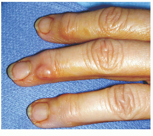
Mucous cysts often present in older adults, usually between fifth and seventh decades, and are commonly associated with osteoarthritis of the distal interphalangeal (DIP) joint. These are rarely painful. Patients are often concerned with cosmesis, or with drainage when these occasionally rupture spontaneously. When the cyst extends toward the eponychial fold, it often causes ridging or deformity of the fingernail. The skin overlying the mucous cyst can become very thin (Fig. 49-3). When these ganglia become symptomatic, the pain is usually related more to underlying osteoarthritis at the DIP joint and less from the mucous cyst.
CLINICAL POINTS
Masses of the finger are common.
Patients typically are young or middle-aged adults.
Ganglia contain thick, mucinous fluid.
PHYSICAL FINDINGS
The clinical presentation of ganglia is classic, and diagnosis can usually be made on the basis of history and physical examination. The hand should be inspected and palpated in standard fashion. A flexor sheath ganglion usually presents as a small, firm, mobile volar mass at the level of the proximal digital flexion crease (Fig. 49-1). It arises from the
A1 pulley and can occasionally be seen in conjunction with a trigger digit. The cyst is attached to the tendon sheath and does not move with the tendon during flexion or extension of the digit. A good neurovascular examination of the finger must be performed to ensure that the mass is not causing any digital nerve compression. The PIP ganglion arises over the dorsolateral aspect of the joint and is relatively fixed in position (Fig. 49-2A).
A1 pulley and can occasionally be seen in conjunction with a trigger digit. The cyst is attached to the tendon sheath and does not move with the tendon during flexion or extension of the digit. A good neurovascular examination of the finger must be performed to ensure that the mass is not causing any digital nerve compression. The PIP ganglion arises over the dorsolateral aspect of the joint and is relatively fixed in position (Fig. 49-2A).
Stay updated, free articles. Join our Telegram channel

Full access? Get Clinical Tree


