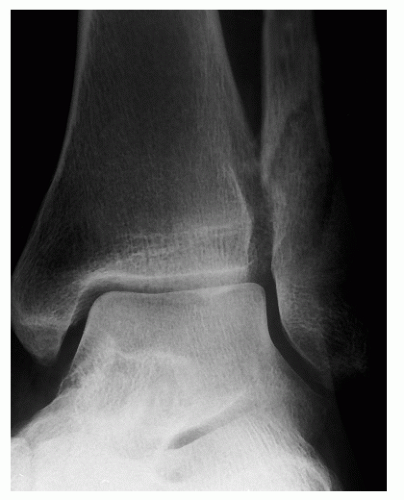
Fractured Fibula (Nondisplaced/Avulsion Fracture)
David I. Pedowitz
CLINICAL PRESENTATION
Ankle fractures are common entities encountered by both primary care physicians and orthopaedic surgeons. They are, in fact, the most common fracture treated by orthopaedic surgeons.1 A fractured fibula, the most common variant of an ankle fracture, generally occurs as the result of a rotational injury to the ankle. They can occur above, below, or at the level of the ankle joint and generally require acute attention as they are quite painful and patients typically cannot ambulate. Avulsion injuries, on the other hand are the separation of only a small piece of extra-articular bone, below the level of the ankle joint. They are due to a rupture of the ligamentous insertion to bone, in which the attachment fails and pulls a small fragment of bone off the fibula with the ligament. Patients typically give the history of a low-energy injury such as twisting and falling on an ankle.2 They will complain of pain and swelling in the area of injury. Furthermore, the patient can have varying levels of discomfort and may or may not be able to ambulate.
Ankle fractures generally manifest in older populations, and their incidence and severity are increasing in patients over the age of 65.3 This may be a consequence of the improved longevity and increased activity observed in older individuals.1 Additionally, obesity and a history of smoking have also been correlated with an increased incidence of ankle fractures.4
Treating physicians should also be aware of other entities that could potentially present acutely with swelling and pain such as a Charcot arthropathy in a patient with diabetic neuropathy.2 As a result, the clinician should always inquire about comorbid conditions such as diabetes, peripheral vascular disease, malnutrition, and tobacco use. It is important to inquire about these factors as they can adversely affect fracture healing.
CLINICAL POINTS
Most nondisplaced ankle fractures do not require surgery.
Most can be allowed to be weightbearing as tollerated in a walking boot.
Diabetics require double the time of immobilization as nondiabetics.
PHYSICAL FINDINGS
Physical examination will reveal swelling and tenderness to palpation at the area of injury. There may be a limitation in range of motion depending on the degree of swelling and pain.
One should also evaluate the circulatory and neurologic status of the extremity to detect if there is underlying vascular disease or neuropathy. Compromise of the neurologic status of the extremity can occur in the presence of a dislocation of the ankle. This is often a stretch injury to a peripheral nerve. These neuropraxias often recover completely over 3 to 6 months. Furthermore, the clinician should also look for any lacerations/abrasions that could indicate that the injury (fracture) communicates with the outside environment (open fracture). Both of these situations are emergent conditions and require evaluation and treatment by an orthopaedic surgeon.2 Proximal leg tenderness with no clearly visible swelling at the level of the ankle is also a cause for concern. This could potentially indicate a high fibular fracture with an injury to the tibiofibular interosseous membrane and syndesmosis (Maisonneuve injury).4
One should also evaluate the circulatory and neurologic status of the extremity to detect if there is underlying vascular disease or neuropathy. Compromise of the neurologic status of the extremity can occur in the presence of a dislocation of the ankle. This is often a stretch injury to a peripheral nerve. These neuropraxias often recover completely over 3 to 6 months. Furthermore, the clinician should also look for any lacerations/abrasions that could indicate that the injury (fracture) communicates with the outside environment (open fracture). Both of these situations are emergent conditions and require evaluation and treatment by an orthopaedic surgeon.2 Proximal leg tenderness with no clearly visible swelling at the level of the ankle is also a cause for concern. This could potentially indicate a high fibular fracture with an injury to the tibiofibular interosseous membrane and syndesmosis (Maisonneuve injury).4
STUDIES (LABS, X-RAYS)
Necessary studies to be obtained when evaluating a suspected fibular fracture should always include radiographs. Standard radiographs include the anteroposterior, mortise, and lateral non-weight-bearing views.4 However, not all injured ankles require radiographs. The Ottawa Ankle Rules were designed to aid health care professionals in deciding on whom to order radiographs to diagnose a suspected ankle fracture.4 The Ottawa Ankle Rules state that radiographs are required to diagnose an ankle fracture if the patient has tenderness near one or both malleoli and has at least one of the following present4:
Stay updated, free articles. Join our Telegram channel

Full access? Get Clinical Tree


