Flatfoot
David A. Spiegel
B. David Horn
CLINICAL PRESENTATION
Flatfoot, or pes planovalgus, is a common finding in infants and children and is rarely a source of discomfort or functional problems. Flatfoot may best be described as a normal variant that is seen in up to 50% of walking-age children1 and 25% of the adult population.2 There may be a positive family history, and flatfeet may be associated with generalized ligamentous laxity (familial), obesity, or a connective tissue disease or syndrome. The longitudinal arch normally reaches its adult height by 10 years of age,3,4 and there is some evidence to suggest that early shoe wear, especially shoes with a stiff sole, may be associated with flatfoot in later life.5 Most flatfeet are flexible and usually cause no symptoms for the patient. Symptoms are more common when a flexible flatfoot is associated with a tendo Achilles contracture6 or when range of motion is restricted (rigid flatfoot), as in a tarsal coalition.
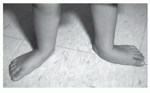
Anatomically, flatfoot may be described as an alteration in the relationship between the bones of the hindfoot, with malalignment at the calcaneo-talo-navicular complex (Fig. 56-1).
The differential diagnosis for flatfoot depends on the age of the patient. Infants and toddlers almost always have the appearance of flatfeet, although both a congenital vertical talus and a calcaneovalgus foot may appear similar to a flexible flatfoot. During the childhood years, the most common diagnosis is a flexible flatfoot, with or without an associated tendo Achilles contracture. In older children and adolescents, the differential diagnosis continues to include flexible flatfeet with associated short tendo Achilles but also includes rigid forms of flatfoot such as tarsal coalition. Flatfoot, either flexible or structural, may also be associated with a variety of other diagnoses, including neurologic diseases (cerebral palsy), syndromes (Down), connective tissue disorders (Marfan, Ehlers-Danlos), or inflammatory diseases such as juvenile rheumatoid arthritis.
CLINICAL POINTS
This common finding in infants and children may occur in adults.
A normal variant in most cases
The patient typically is asymptomatic.
Pathology involves malalignment of the bones of the hindfoot.
PHYSICAL FINDINGS
The musculoskeletal examination focuses on alignment and range of motion and should include an assessment of the degree of soft tissue laxity and a careful neurologic examination. An examination of the hips should be performed, especially in infants with a foot deformity as there is a reported association between foot deformity in infants and developmental dysplasia of the hip. A flexible flatfoot will have a normal appearance in the non-weight-bearing position; however, the arch will flatten or disappear when the patient is standing (Fig. 56-1). The skin over the medial hindfoot should be inspected for calluses or signs of irritation.
In infants, a calcaneovalgus foot may be confused with a flatfoot (Fig. 56-2). The calcaneovalgus foot will be everted and dorsiflexed, and the dorsum of the foot will often touch the lateral surface of the lower leg. The relationship between the midfoot and the hindfoot will be normal, and there is excessive dorsiflexion through the ankle.
Stay updated, free articles. Join our Telegram channel

Full access? Get Clinical Tree


