External Fixation Postoperative Care and Rehabilitation
Paul S. Cooper
Introduction
Complications seen in the postoperative period with the use of external fixation in the lower extremity are numerous and range from mild to severe. While the most common complication is soft tissue irritation and/or infection around the wire/pin site, this is not the most drastic. More severe complications involve contractures, axial deviation of the lower extremity, severe swelling, nerve injury, potential refracture, loss of correction, or deep vein thrombosis with possible extension to a pulmonary embolus.
Immediate Postoperative Period
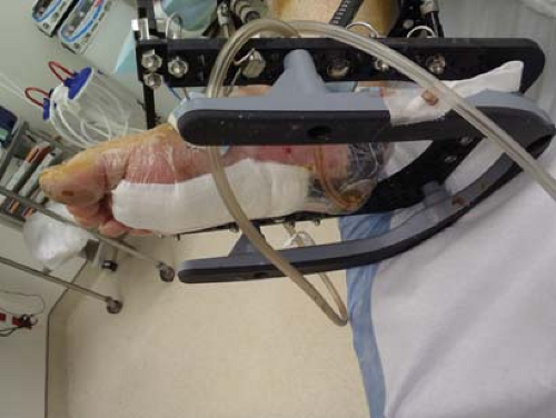
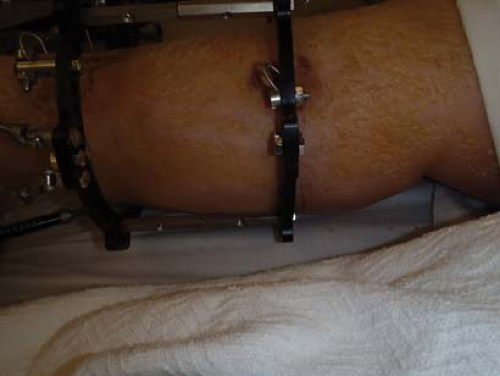
Primary issues in the immediate postoperative period while still in the hospital setting include wound and wire/pin care, appropriate antibiosis, pain management, and anticoagulation considerations. Generally, the postoperative dressings applied for both the incisions and wires/pins remain unchanged until the first follow-up visit in the clinical setting. Patients with active wounds may have a negative pressure wound therapy (NPWT) applied intraoperatively. Typically, a NPWT device may lose the suction seal due to the wire/pin density around the wound and should be checked daily for integrity (Figure 23.1). The risk for deep vein thrombosis stems from a lack of muscle action in the lower extremity, especially when the external fixator spans one or more major joints. Children and most healthy adults do not usually require anticoagulation while in the external fixation device. High-risk candidates for anticoagulation therapy postoperatively include those with coagulopathy disorders, prior deep vein thrombosis or pulmonary emboli, and certain neoplastic disorders. Elastic wraps may be also used on the lower extremity involved to add some degree of compression and keep any potential edema formation in check. On rare occasions when the patient’s swelling places the lower extremity at risk for impingement on the external fixation rings, a trial course of diuretics may be also indicated (Figure 23.2). While in the hospital setting, the patient should have daily physical therapy to work on uninvolved adjacent joint motion and independence with non–weight bearing gait training using a walker, crutches, or other assistant devices. In addition, patients should refrain from smoking due to the higher rate of soft tissue and osseous complications following application of the external fixation device.
Hospital Discharge
Following patient discharge, the first return visit to the outpatient clinic may occur within 10 to 14 days. During this visit and subsequent visits, standard foot, ankle, and lower extremity radiographs should be obtained for evaluation of the index procedure. In cases where either lower limb lengthening or compression arthrodesis occurs, radiographs of the adjacent uninvolved joints should similarly be included in the series to evaluate for potential joint incongruency. Patients may follow up every 1 to 2 weeks as needed during the active external fixation adjustment phase. In cases where a static circular external fixator was utilized and no postoperative corrections through the external fixator are occurring, visits may be spaced to every 3 to 4 weeks. In cases of arthrodesis or lower extremity
lengthening, an external bone stimulator may be applied to help facilitate the healing process. Several units have been made to be attached directly to the external fixator through an adaptor if needed during the postoperative period.
lengthening, an external bone stimulator may be applied to help facilitate the healing process. Several units have been made to be attached directly to the external fixator through an adaptor if needed during the postoperative period.
Postoperative Clinic Visits
Wire/pin tract issues are the greatest potential complication and challenge following application of external fixators. Wire/pin vulnerability for irritation or infection is dependent on location, position of the transosseous wire or half-pin, the degree of soft tissue overlying the wire/pin site, external fixation stability, and risk for trauma during the postoperative period. In the immediate perioperative period, petroleum gauze wrapped around the pin sites combined with wire/pin sponges or Betadine-soaked adaptic dressings applied at the time of surgery meet the minimum needs. In the postoperative period, the initial wire/pin dressing needs to be inspected and cleaned and wire/pin sites may be addressed with normal saline alone, antibacterial soap and water, or Betadine-soaked non-adherent dressings. Irritants such as alcohol or peroxide should be avoided for risk of inflaming surrounding soft tissue. A bacterial ointment may be applied at the wire/pin interface followed by application of the dressing. Commercially available wire/pin sponges are now available that have silver-impregnated antibacterial properties which can be applied with a wire/pin clamp to minimize soft tissue excursion (see Chapter 5). Alternatively, a cut 2 × 2 in. or 4 × 4 in. in multiple layers combined with a Kerlix wrap allows minimal soft tissue excursion around the wires/pins, thus reducing risk for irritation or infection (see Chapter 5). Crusting around the wires/pins may develop and by itself is not a sign or increased risk for infection. The wire/pin crusts can be removed safely thus reducing the potential for trapping bacteria underneath the soft tissue crust (Figure 23.3). Wire/pins may be noted to demonstrate some redness in a zone around the wire/pin which blanches under pressure. This case is more related to wire/pin inflammation and a nonsteroidal anti-inflammatory may help with addressing discomfort associated with inflammation signs (Figure 23.4). Wire/pin irritation may increase as additional wire/pin tension develops in cases of compression or distraction osteogenesis. In these cases, the corresponding skin in front of the wire/pin may need to be released with a no. 11 blade (Figure 23.5). This technique can be performed under local anesthesia in the clinical setting when warranted.
Stay updated, free articles. Join our Telegram channel

Full access? Get Clinical Tree


