Electrodiagnostic Studies


Electrodiagnostic Studies


Electrodiagnostic studies (also known as NCS/EMG or sometimes just EMG) include nerve conduction studies (NCSs or NCVs) and EMG. Other less commonly performed electrodiagnostic tests include somatosensory evoked potentials, brainstem auditory evoked potentials or responses, single-fiber EMG (SFEMG), repetitive stimulation studies, and sympathetic skin response. This discussion is limited to the most commonly used studies, NCS and EMG. As they are usually performed together and reported as one comprehensive report, they will be referred to as a single test (NCS/EMG). This test provides physiologic information about nerves and muscles in real time. It gives information about muscle and nerve function, unlike most radiologic studies, which give a static picture of anatomy and do not directly assess function.

Indications for electrodiagnostic testing include numbness, tingling/paresthesias, pain, weakness, atrophy, depressed deep tendon reflexes, and/or fatigue. EMG/NCS can serve as an important part of a patient’s clinical picture. Electrodiagnostic tests are used to (1) establish a correct diagnosis, (2) localize a lesion, (3) determine the treatment when a diagnosis is already known, and (4) provide information about the prognosis.1 NCS/EMG should be considered an extension of a good history and physical examination.


INITIAL SETTINGS FOR NCS

Sweep speed is the horizontal axis on the recording in units of time (milliseconds [ms]). Gain is the vertical axis on the graph in units of voltage (millivolts [mV] for motor studies or microvolts [μV] for sensory studies).

Motor settings: sweep – 2 ms/division, gain – 5 mV/division.

Sensory settings: sweep – 2 ms/division, gain – 20 μV/division.1


INITIAL SETTINGS FOR EMG

Sweep speed: 10 ms/division

Low-frequency filter: 10 to 30 Hz

High-frequency filter: 10,000 to 20,000 Hz

Amplifier sensitivity: 50 to 100 μV1


INTRODUCTION TO NCS

NCS is the recording of an electrical response of a nerve (via an electrode over that nerve or a muscle) that is stimulated (electrically depolarized using a probe) at one or more sites along its course. The action potential (AP) that is propagated is the summative response of many individual axons or muscle fibers. For motor nerves, this response is called a compound motor action potential (CMAP) and represents the summative response of motor
units (MUs) that are firing. CMAPs are usually recorded in mV. For sensory nerves, the response is called an SNAP and represents the summation of individual sensory nerve fibers. SNAPs are very small-amplitude potentials that are usually recorded in μV. Late responses (evoked potentials that record over a very long pathway) include F waves and H-reflexes. Orthodromic refers to conduction in the same direction as occurs physiologically (i.e., a sensory fiber conducts from the extremity toward the spine). Antidromic refers to conduction in the opposite direction to the physiological direction.


Components of the AP

Latency is the time it takes from stimulation to the beginning of the AP (the speed of transmission). The latency of a sensory nerve is dependent on the conduction speed of the fastest fibers and the distance it travels. The latency of a motor nerve also includes the time it takes for the AP to synapse at the NMJ and the speed of conduction of the electrical potential through the muscle. Since there is no myoneural junction of sensory nerves, the latency of a sensory nerve is directly related to the conduction velocity (CV). Latency measurement requires standardized and accurately recorded distance or else the results are meaningless.

Conduction velocity reflects how fast the nerve AP is propagating. In sensory studies, the velocity is measured directly from the time it takes the AP to travel the measured distance (distance/latency). In a motor nerve, two different sites have to be stimulated to calculate the velocity (velocity = change in distance/change in time) and account for the myoneural junction. The presence of a myelin covering speeds up NCV via a process known as saltatory conduction. Myelinated nerves conduct impulses approximately 50 times faster than unmyelinated nerves. In myelinated nerves, the CV is primarily dependent on the integrity of the myelin covering. Slowing or latency prolongation usually implies demyelination.

Amplitude correlates with axonal integrity. Decreased amplitude could indicate an axonal lesion (if the amplitude is decreased both distally and proximally) or it can indicate a conduction block across the site of injury (if the amplitude is low distally and not proximally).2,3


TYPES OF NERVE INJURIES

Nerve injuries can be classified depending on whether there is injury to the axon, the myelin, or both. Often, especially with trauma, the affected structures do not always fit into one category. It is the job of the electromyographer to diagnose and communicate the type of injury that exists, the severity, and the location. Seddon proposed a classification of nerve injuries in 1943 that is still commonly used as it correlates well with electrophysiology:

1. Neurapraxia – defined as conduction block. This type of nerve injury occurs in the peripheral nerve with minor contusion or compression. There is preservation of the axon; only the myelin is affected. The transmission of APs is interrupted for a brief period, but recovery is usually complete in days to weeks.

2. Axonotmesis – more significant injury: breakdown of axon with accompanying Wallerian degeneration distal to the lesion. There is preservation
of some of the supporting connective tissue stroma (Schwann cells and endoneurial tubes). Regeneration of axons (through collateral sprouting or axonal growth) can occur with good functional recovery, depending on the amount of axonal loss.

3. Neurotmesis – severe injury with complete severance of the nerve and its supporting structures; extensive avulsing or crush injury. The myelin, axon, perineurium, and epineurium are all disrupted. Spontaneous recovery is not expected.

Injury to the myelin can be focal (local), uniform (throughout the nerve), or segmental (affecting some parts of the nerve but not others):


  • Uniform demyelination – slowing of CV along the entire nerve (e.g., Charcot-Marie-Tooth disease).


  • Segmental demyelination – uneven degree of demyelination in different areas along the course of the nerve; may have variable slowing (temporal dispersion).


  • Focal nerve slowing – localized area of demyelination causing nerve slowing; decreased CV is noted across the lesion.


  • Conduction block – severe focal demyelination that prevents propagation of the AP through the area. There will be more than 20% amplitude decrement when the nerve is stimulated proximal to the lesion. The distal CMAP amplitude remains intact. Clinically, conduction block presents as weakness.

Axonal injuries will lead to Wallerian degeneration distal to the lesion. Low-amplitude CMAPs will be noted with both proximal and distal stimulation. On EMG, abnormal spontaneous potentials (fibrillations [fibs] and positive sharp waves [PSWs]) are seen. The MU recruitment will be decreased (increased firing frequency of existing MUs). With reinnervation, MUs may become polyphasic with high amplitude and long duration.4,5


H-REFLEX

The H-reflex (Hoffmann reflex) is a true reflex and is the electrical equivalent of the monosynaptic or oligosynaptic stretch reflex. It is a sensitive but nonspecific tool for possible S1 radiculopathy, especially when clinical, radiologic, and electrophysiologic signs of motor root involvement are lacking. In some cases, it may be the only abnormal study. The H-reflex is usually elicited by submaximally stimulating the tibial nerve in the popliteal fossa. Such stimulation can be initiated by using slow (less than 1 pulse/s), long-duration (0.5 to 1 ms) stimuli with gradually increasing stimulation strength. The stimulus will travel along the most excitable Ia afferent nerve fibers, through the dorsal root ganglion (DRG). It then gets transmitted across the central synapse to the anterior horn cell, which then sends it down along the alpha motor axon to the muscle. Hence, the H-reflex is a measure of the time it takes for the orthodromic sensory response to get to the spinal cord proximally and the orthodromic motor response to reach the muscle distally (on which the recording electrode is placed). A generally acceptable result would be a motor response usually between 0.5 and 5 mV in amplitude and a latency of 28 to 30 ms. H-reflex studies are usually performed bilaterally because asymmetry of responses is an important
criterion for abnormality. An abnormal latency greater than 0.5 to 1.0 ms (as compared with the other side) or H-reflex absence in patients under 60 years may suggest a lesion along the H-reflex pathway (afferent and/or efferent fibers). This may be due to an S1 radiculopathy. The standard formula for calculating the H-reflex is 9.14 + 0.46 (leg length in cm from the medial malleolus to the popliteal fossa) + 0.1 (age). For a patient older than 60 years, 1.8 ms will be added to the total calculated value.

In normal infants or adults with UMN (corticospinal tract) lesions, the H-reflex may be elicited in muscles other than the gastrocnemius/soleus muscles or flexor carpi radialis. It is often absent in patients older than 60 years. The reflex can be potentially inhibited by antagonist muscle contractions and initiated by agonist muscle contractions.

The H-reflex does have some limitations. It is unable to distinguish between acute and chronic lesions, may be normal with incomplete lesions, is diluted by focal lesions, and is nonspecific in terms of injury location. Once the H-reflex is found to be abnormal, it will usually remain so, even with resolution of symptoms.


F WAVES

F wave or F response is a small-amplitude, variable-latency late motor response that occurs following the activation of motor nerves. It derives its name from the word “foot” because it was first recorded from the intrinsic foot muscles. Unlike the H-reflex, the F wave does not represent a true reflex because there is no synapse from an afferent impulse to a motor nerve. Depolarizing peripheral nerves with external stimuli evokes potentials propagating both proximally and distally. Electrical stimulation of a peripheral nerve results in an orthodromic CMAP. In addition, the proximally (antidromically) propagating potential activates a small percentage of anterior horn motor neurons. In turn, this generates an orthodromic motor response (the F wave) along the same axon that activates a few muscle fibers picked up by the recording electrode.6,7

F waves can be obtained from any muscle by a supramaximal stimulus. Because of their variability (as opposed to H-reflexes), multiple stimulations must be used to obtain the shortest latency. F waves may be useful in the evaluation of peripheral neuropathies with predominantly proximal involvement, such as Guillain-Barré syndrome and chronic inflammatory demyelinating polyneuropathies, in which distal conduction velocities may be normal early in the disease. However, the value of the F wave in evaluating focal nerve lesions, such as radiculopathy or peripheral nerve entrapment, is extremely limited largely due to the variability of F-wave responses. In addition, most muscles receive innervation from multiple roots, so the fastest (nonaffected) fibers will be normal, as well as the fact that the results are nonspecific. It is a pure motor response, and its long neural pathway dilutes focal lesions and hinders the specificity of injury location. F waves are also generally not seen in nerves where the CMAP amplitude is severely reduced, such as severe axonal loss, since the F-wave amplitude is only 1% to 5% of the amplitude of the CMAP.

Normal latency of F wave: upper limb: 28 ms; lower limb: 56 ms. Side-toside difference: <2.0 ms for upper limbs; <4.0 ms for lower limbs.


Blink Reflex

The most complicated of the late responses is the blink reflex. It is the electrophysiologic correlate of the corneal reflex. The sensory afferent limb of the reflex is the supraorbital nerve, a branch of the ophthalmic division of the trigeminal nerve (CN V1). Intervening synapses (pons and medulla) are stimulated. The motor efferent limb is the facial nerve (CN VII), which innervates the orbicularis oculi muscle. As with the corneal reflex, stimulation of one side of the supraorbital branch of the trigeminal nerve elicits a motor response (eye blink) bilaterally through the facial nerves. Abnormalities anywhere along the reflex arc (central or peripheral) can be detected.

There is an early response (R1) due to a disynaptic reflex arc from the ipsilateral sensory nucleus of V to the ipsilateral facial nerve. There is also a late response (R2) due to multiple interneurons connecting the ipsilateral sensory nucleus of V to the ipsilateral spinal motor nucleus of V and then to the bilateral facial nuclei.

Recording electrodes are placed below and slightly lateral to the pupils bilaterally. Reference electrodes are placed just lateral to the lateral canthus bilaterally. The ground can be placed on the chin. The stimulator is placed over the medial supraorbital ridge of the eyebrow. The sweep speed should be 5 or 10 ms with initial sensitivity of 100 or 200 μV.

Normal latency for R1 response is <13 ms. Normal latency for ipsilateral R2 is <41 ms. Normal latency for contralateral R2 is 44 ms. Acceptable normal bilateral variation for R1 is <1.2 ms, for ipsilateral R2 is <5 ms, and for contralateral R2 is <7 ms (Table 17-1).


TABLE 17-1 Basic Abnormal Patternsa


Lesion


Electrodiagnostic pattern if affected side stimulated


Electrodiagnostic pattern if unaffected side stimulated


Unilateral CN V


Delayed (partial injury) or absent (complete injury) R1 and bilateral R2


Normal R1 and bilateral R2


Unilateral CN VII


Delayed or absent R1 and ipsilateral R2


Delayed or absent contralateral R2


Unilateral midpontine


Delayed R1 and normal bilateral R2


Normal R1 and bilateral R2


Unilateral medullary


Delayed ipsilateral R2


Delayed contralateral R2


Demyelinating peripheral neuropathy


Possible delay or absence of R1 and bilateral R2


Possible delay or absence of R1 and bilateral R2


a Using the anatomy outlined above and these basic patterns, complex and bilateral lesions can be extrapolated.


Preston DC, Shapiro BE. Blink reflex. In: Electromyography and Neuromuscular Disorders. 2nd ed. Philadelphia, PA: Elsevier; 2005:59-64.


EMG INCLUDING MONOPOLAR VERSUS CONCENTRIC NEEDLE

EMG testing involves evaluation of the electrical activity of skeletal or voluntary muscles. Muscles contract and produce movement through the orderly recruitment of MUs. An MU is defined as one anterior horn cell, its axon, and all the muscle fibers innervated by that motor neuron. An MU is the fundamental structure that is assessed in EMG testing. EMG requires a thorough knowledge of the anatomy of the muscle being tested in order to place the needle electrode in the appropriate muscle.

Monopolar needles are 22G to 30G Teflon-coated stainless steel needles with an exposed tip of 0.15 to 0.2 mm2 (Fig. 17-1A). They require a surface electrode or a second needle as a reference lead. Another surface electrode serves as a ground. A monopolar needle records the voltage changes between the tip of the electrode and the reference. Since it picks up from a full 360° field around the needle, it registers larger amplitude and has increased polyphasicity when compared with the concentric needle. The smaller diameter and the Teflon coat make the monopolar needle less uncomfortable. This, combined with its cost advantage over the concentric, has led to its preferential clinical use.

Concentric needles are 24G to 26G stainless steel needles (Fig. 17-1B). The needle comprises a reference (cannula) electrode with a bare inner
wire in the center of the shaft that is the recording electrode. The concentric needle can register the voltage changes between the wire and the shaft. The pointed tip of the needle has an oval (beveled) shape. Since the exposed active recording electrode is on the beveled portion of the cannula, the concentric needle picks up from a 180° field. Therefore, it registers smaller amplitude (since it has a smaller recording area). A separate surface electrode serves as the ground.


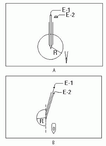
Figure 17-1 Needle electrodes. A. Monopolar needle. B. Concentric needle.

Adapted from Dumitru D, ed. Electrodiagnostic Medicine. 2nd ed. Philadelphia, PA: Hanley & Belfus; 2002.


EFFECTS OF TEMPERATURE AND AGE ON NCS

Cooling is thought to prolong the opening of Na+ channels.8,9 Decreasing the temperature of a limb affects SNAPs and CMAPs by prolonging latency, decreasing CV, and increasing amplitude and duration. CV decreases 2.4 m/s per 1° C decrease. Correction formulas exist but the best approach is to warm the limb prior to the NCS (32° in the upper limbs and 30° in the lower limbs).

As patients age, SNAP and CMAP amplitudes decrease and latencies increase. Motor NCSs for newborns are 50% of adult values since myelination is incomplete. Normal adult values are attained by age 5 years. After age 60 years, there is a progressive decline of 1 to 2 m/s per decade in the NCS of the fastest motor fibers.


THE NEEDLE EMG EXAMINATION

The EMG evaluation typically has four components:

1. Insertional activity

2. Activity at rest

3. MU analysis

4. Recruitment


Insertional Activity

Healthy muscle is electrically silent at rest. Insertional activity8,9 refers to the brief electrical activity associated with the needle entering the sarcolemma, which causes muscle fiber injury. The associated sound should be crisp. Insertional activity is classified in three ways:


  • Normal insertional activity only lasts a few hundred milliseconds and is due to muscle depolarization.


  • Increased insertional activity occurs due to denervation or cell membrane irritability and lasts >300 ms. There may be evidence of initially positive deflection waveforms that do not persist. If these positive waveforms are sustained and fire regularly, they are considered abnormal spontaneous potentials (see below).


  • Decreased insertional activity occurs when the needle is placed into atrophied muscle, fat, or edema and lasts <300 m/s.


Activity at Rest


NORMAL SPONTANEOUS ACTIVITY

A needle should be inserted into a muscle at three to four different depths and in three to four different directions (examining three or four
electrically discrete areas of muscle) for insertional activity and activity at rest. The needle can be withdrawn almost to the skin and then redirected in a different direction, again stopping at three or four different depths. This can be repeated so that the needle examines about 12 to 16 discrete areas of the muscle (depending on the patient’s tolerance).


  • After insertion of the needle into normal muscle at rest, there should be electrical silence (Fig. 17-2A).


  • Normal muscle may also display end plate activity. This occurs after a needle is placed in the region of the NMJ or end plate. The needle should be moved out of the end plate, as the clinician cannot get reliable information about the muscle. Either of two waveforms may occur: miniature end plate potentials (MEPPs) or end plate potentials (EPPs). The patient may complain of increased pain. It is important to recognize these potentials so that they are not misinterpreted as abnormal spontaneous potentials.


    • MEPPs – Represent spontaneous release of single quantum of acetylcholine (ACh) at the presynaptic terminal that manifests as end plate noise (Fig. 17-2B).


    • EPPs or “end plate spikes” – Represent single muscle fiber depolarizations at the presynaptic terminal with resultant release of large amounts of ACh (Fig. 17-2C).


  • MEPPs and EPPs may or may not be present together (Fig. 17-2D).


ABNORMAL SPONTANEOUS ACTIVITY


  • Usually represents pathology (injury or denervation) that stems from a muscle or nerve. These spontaneous depolarizations have an abnormal morphology and firing pattern.


  • Examples of muscle fiber-generated spontaneous potentials: Fibs triphasic with initial positive (downward) deflection, PSWs biphasic with positive deflection, myotonic discharges, and complex repetitive discharges (CRDs).


    Figure 17-2 End plate activity.

    Adapted from Dumitru D, ed. Electrodiagnostic Medicine. 2nd ed. Philadelphia, PA: Hanley & Belfus; 2002.

    Courtesy of DeLisa JA, ed. Manual of Nerve Conduction Velocity and Clinical Neurophysiology. 3rd ed. New York, NY: Raven Press; 1994.


  • Examples of neural-generated spontaneous potentials: Myokymic discharges, cramps, neuromyotonic discharges, tremors, fasciculations, and multiple MU potentials.


  • Fibs and PSWs appear 3 weeks or more after injury.


  • Abnormal spontaneous potentials are usually of small amplitude. Therefore, the gain on the EMG machine should be set to 50 to 100 μV for the best visualization (Fig. 17-3).

Grading of fibs and PSWs is from 0 to 4+, with a sweep of 10 ms/division

(0) no fibs or PSWs present

(1+) one fib/PSW per screen persistent within two areas

(2+) fibs/PSWs in greater than two areas, about two per screen

(3+) fibs/PSWs in most muscle regions, greater than half of the screen

(4+) fibs/PSWs in all areas of the muscle and fill the entire screen

Fasciculation potentials originate from a single MU and may have an intermittent or a normal firing pattern. When associated with PSWs or fibs, they suggest pathology. In the absence of fibs or PSWs, they may be due to stress, fatigue, or caffeine (Fig. 17-4).


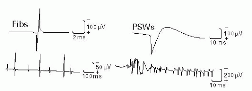
Figure 17-3 Fibs and PSWs.

Adapted from DeLisa JA, ed. Manual of Nerve Conduction Velocity and Clinical Neurophysiology. 3rd ed. Baltimore, MD: Raven Press; 1994.


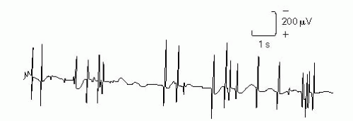
Figure 17-4 Fasciculation potentials.

Adapted from DeLisa JA, ed. Manual of Nerve Conduction Velocity and Clinical Neurophysiology. 3rd ed. Baltimore, MD: Raven Press; 1994.


CRDs frequently result from denervation and reinnervation through collateral sprouting. Their presence suggests a chronic process such as chronic radiculopathy, peripheral neuropathy, anterior horn disease, polymyositis, or myxedema (Fig. 17-5).

Myotonic discharges (Fig. 17-6) originate in the muscle due to membrane instability. They have a characteristic waxing and waning character and have been compared with a dive-bomber sound. They are commonly seen in myotonic dystrophy, myotonia congenita, polymyositis, chronic radiculopathy, peripheral neuropathy, maltase deficiency, and hyperkalemic periodic paralysis (Table 17-2).


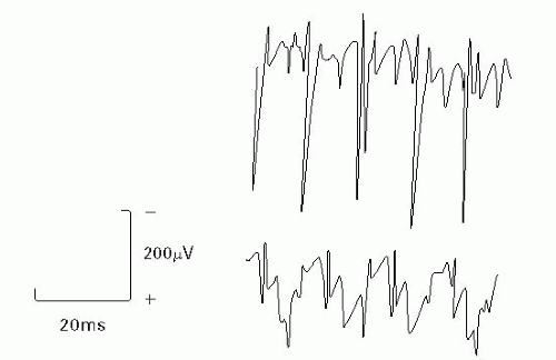
Figure 17-5 CRDs.

Adapted from DeLisa JA, ed. Manual of Nerve Conduction Velocity and Clinical Neurophysiology. 3rd ed. Baltimore, MD: Raven Press; 1994.


Figure 17-6 Myotonic discharges.

Adapted from Goodgold J. Electrodiagnosis of Neuromuscular Diseases. 2nd ed. Baltimore, MD: Williams & Wilkins; 1977.


TABLE 17-2 Abnormal Spontaneous Potentials


MMEPs


EPPs


Fib potentials


Positive sharp waves


Fasciculation potentials


Complex repetitive discharge


Myotonic discharges


Sound


Sea shells


Sputtering fat in a hot pan


Drops of rain on a tin roof


Dull thud


Varies


Misfired motor boat


Dive bomber


Firing pattern


Irregular


Irregular


Regular


Regular


Irregular


Regular/starts and stops abruptly


Waxes and wanes


Duration (ms)


1-2


3-5


1-5


10-30


5-15


Variable


> 5-20


Amplitude (μV)


10-20


100-200


20-1,000


20-1,000


> 300 m


50-500


20-300


Rate (Hz)


150


50-100


0.5-15


0.5-15


0.1-10


10-100


20-100


Waveform/deflection


Monophasic negative (upward)


Biphasic negative (upward)


Triphasic with initial positive (downward)


deflection Biphasic with initial positive (downward) deflection


Similar to motor unit action potential (MUAP)


Similar to MUAP, fibs, and PSWs


Similar to EPP, fibs, and PSWs


Cause


MEPPs


Irregularly firing muscle fiber APs


Spontaneous depolarization of a muscle fiber


Spontaneous depolarization of a muscle fiber


Spontaneous involuntary discharge of single MU


Depolarization of single muscle fiber with ephaptic spread to adjacent denervated fibers


Spontaneous discharge of a muscle fiber


Seen in


Needle in the end plate


Needle in the end plate


Denervation (may be due to neurogenic muscle disorder or disorders of the NMJ )


Denervation (may be due to neurogenic, muscle disorder or disorders of the NMJ )


Processes that affect the lower motor neuron (LMN). Also seen in benign fasciculations


Chronic neuropathic and myopathic disorders


Myotonic dystrophy, myotonia congenita and paramyotonia, some myopathies, hyperkalemic periodic paralysis, and rarely in denervation


Other potentials that are nerve generated include cramp potentials, myokymia, and neuromyotonia. These are beyond the scope of our discussion here. Adapted from Preston DC, Barbara ES. Electromyography and Neuromuscular Disorders: Clinical-Electrophysiologic Correlations. Philadelphia, PA: Butterworth-Heinemann; 2005:215-225.


MU Analysis

After analysis of insertional activity and spontaneous activity, the next step is to analyze the motor unit action potentials (MUAPs). First assess the morphology (duration, amplitude, phases, and rise time). This should be done during a minimal contraction (sometimes positioning changes can bring this on). A trigger and delay line can be helpful in assessing MU stability.

An MU is defined as an individual motor neuron, the muscle fibers it innervates (ranging from five to hundreds), and the NMJs between these two components. MUAP morphology varies depending on the age of the patient and the muscle being tested.

Duration is measured from the initial deflection from the baseline to the return to baseline (typically 5 to 15 ms) and reflects the synchrony of muscle fibers firing. The duration is increased with asynchronous firing of the fibers of an MU (as in reinnervation or other neuropathic processes) and is decreased in myopathic processes (fewer fibers contribute to the MU). When listening to the MUs, duration correlates with pitch; thus, long duration is dull and thuddy and short duration is crisp and staticlike.

Amplitude is measured from the most positive to the most negative peak of the MU and reflects fiber density. The criteria for normal amplitude depend on the type of needle used (several hundred microvolts to a few millivolts for concentric needles, 1 to 7 μV for a monopolar needle). Amplitude increases (1) as the needle approximates the MU, (2) as the number of muscle fibers of the MU is increased, (3) with increasing diameter of the muscle fibers (muscle fiber hypertrophy), and (4) with more synchronous firing of the muscle fibers. Also, amplitude may be increased after reinnervation (neuropathic injuries) and may be decreased in myopathies. When listening to the MUs, amplitude correlates with volume (not pitch).

Phases are determined by counting the number of baseline crossings and adding one. Polyphasia implies asynchronous firing of muscle fibers within an MU. Polyphasia is nonspecific and can be seen in both neuropathic and myopathic lesions. The MUAP is generally 2 to 4 phases. All muscles will normally exhibit about 10% polyphasia except the deltoid (up to 25% is normal). When listening to the MUs, polyphasia results in a clicking sound.

Only gold members can continue reading. Log In or Register to continue

Stay updated, free articles. Join our Telegram channel

Jun 19, 2016 | Posted by in PHYSICAL MEDICINE & REHABILITATION | Comments Off on Electrodiagnostic Studies

Full access? Get Clinical Tree

Get Clinical Tree app for offline access