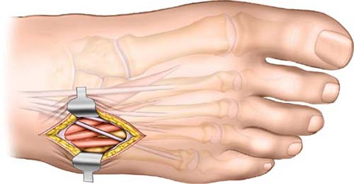
 Dorsolateral Approach to Lisfranc’s Joint
The dorsolateral approach to the lateral part of Lisfranc’s joint is often used in conjunction with the medial approach. In this area, full-thickness skin flaps must be made without undermining any soft tissue. This is most important if two incisions are used (dorsomedial and dorsolateral). The lateral side of the midfoot is mobile and in cases of fractures is frequently stabilized on a temporary basis. The medial side of the midfoot provides stability. Treatment of this part of the joint in cases of fracture often involves fusion.
Position of the Patient
Place the patient supine on the operating room table (see Fig. 7-1). Place a sand bag underneath the buttock of the affected side to correct the natural external rotation of the leg. This maneuver will position the foot for both open and closed procedures, when fluoroscopy is used. Exsanguinate the leg, then apply a tourniquet to the middle of the thigh.
Landmarks and Incision
Although you can palpate the styloid process of the fifth metatarsal laterally and the dorsal surface of the fourth metatarsal, fluoroscopy is necessary for precise anatomic localization of the small bones of the midfoot.
Make a 2- to 4-cm longitudinal incision directly over the dorsal aspect of the fourth metatarsal (Fig. 31-1). The incision may need to be positioned more medially or more laterally depending on the pathology to be treated and the technique to be used. An incision over the fourth metatarsal will allow easy access to the joints between the bases of the fourth and fifth metatarsal and the cuboid as well as the joint between the base of the third metatarsal and the lateral cuneiform.
Stay updated, free articles. Join our Telegram channel

Full access? Get Clinical Tree


