 Dorsal Approaches to the Middle Part of the Foot
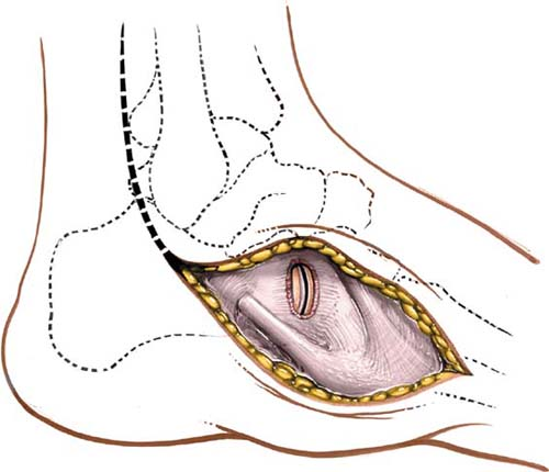
The middle part of the foot extends from the calcaneocuboid and talonavicular joints to the tarsometatarsal Lisfranc’s joints. All these bones and joints are superficial and can be approached directly by dorsal, medial, lateral, and plantar approaches. Operations in this area (which are performed rarely) usually involve surgery on the insertions of the four powerful muscles that, together, are responsible for controlling inversion and eversion of the foot. These muscles are the tibialis anterior, which inserts into the medial surface and undersurface of the medial cuneiform bone, and into the adjoining part of the base of the first metatarsal bone; the peroneus longus, which inserts into the lateral side of the medial cuneiform bone; the peroneus brevis, which inserts into the base of the lateral side of the fifth metatarsal bone; and the tibialis posterior, which inserts into the tuberosity of the navicular bone, the inferior surface of the medial cuneiform bone, the intermediate cuneiform bone, and the bases of the second, third, and fourth metatarsal bones (see Figs. 25-2, 25-5, and 25-9).
The middle part of the foot is the target of various specialized procedures for the treatment of muscle imbalance, mobile flatfoot, and an accessory navicular bone. It is also approached for open reduction and internal fixation of fractures in and around Lisfranc’s joint, and for local tarsal fusion. Only the general surgical approaches are considered here, because the details of operative technique and indications are beyond the scope of this book.
Position of the Patient
Place the patient supine on the operating table. Dorsomedial approaches and medial approaches are carried out with the leg in its natural position of slight external rotation, whereas dorsolateral approaches require internal rotation of the limb, which is achieved by placing a sandbag under the buttock. For all procedures, exsanguinate the limb either by elevating it for 3 to 5 minutes or by applying a soft rubber bandage. Then, inflate a tourniquet (see Fig. 7-1).
Landmarks and Incisions
Landmarks
To palpate the first metatarsal cuneiform joint, feel along the medial border of the foot in a distal to proximal direction. The first metatarsal flares slightly at its base to meet the first cuneiform.
Continue moving proximally along the medial border of the foot to reach the tubercle of the navicular.
The medial side of the talar head is immediately proximal to the navicular. It can be located by inverting and everting the forepart of the foot. The motion that occurs between the talus and the navicular is palpable (Fig. 34-1).
Related posts:

Stay updated, free articles. Join our Telegram channel

Full access? Get Clinical Tree


