Fractures involving the distal radius and ulna are commonly seen in children and adolescents. Management of these injuries in pediatric patients should include assessment of the neurovascular status of the extremity, associated soft-tissue injury, and, most importantly, possible involvement of the physes of the radius and ulna. Treatment of these injuries may vary from simple casting and radiographic follow-up to urgent reduction and surgical fixation. Regardless of the initial treatment plan, the treating surgeon must remain aware of the potential for both early and late complications that may affect outcomes.
Key points
- •
Fractures involving the distal radius-ulna are among the most common fractures seen in the pediatric population.
- •
Distal radius fractures most often result from a fall onto the outstretched hand. An increasing incidence may be related to trends in leisure/sports activities.
- •
The vast majority of these fractures may be treated appropriately with closed reduction and casting.
- •
The clinician should be aware of potential complications such as acute carpal tunnel syndrome, growth arrest and malunion.
Introduction
Fractures involving the distal radius and ulna are commonly seen in children and adolescents. Management of these injuries in pediatric patients should include assessment of the neurovascular status of the extremity, associated soft-tissue injury, and, most importantly, possible involvement of the physes of the radius and ulna. Treatment of these injuries may vary from simple casting and radiographic follow-up to urgent reduction and surgical fixation. Regardless of the initial treatment plan, the treating surgeon must remain aware of the potential for both early and late complications that may affect outcomes. The clinician often must balance the patient and family’s desire for early return to activity with the goal of long-term functionality of the involved limb. Many studies have discussed optimal treatment methods with regards to specific fracture patterns. Nonetheless, management of these injuries tends to differ quite significantly among clinicians. Recently published data have questioned long-held principles of nonoperative management for all fractures. This article reviews distal pediatric forearm fracture management with emphasis on potential complications and discussion related to recently published clinical data.
Epidemiology
Fractures in the pediatric population are common. An annual fracture incidence of 180 per 10,000 in children younger than 16 years has been reported. Fractures of the distal radius were found to be the most common, representing 31% of all fractures in this patient population and tended to occur in the nondominant extremity in roughly 53% of cases. The mean age at the time of fracture was 9.3 years in girls and 10.4 years in boys. Pediatric fractures are more commonly seen in boys, with a male to female incidence ratio of 1.5.
Distal radius fractures most often occur as a result of a fall onto the outstretched hand. Randsborg and colleagues reported that activity-related fracture was most common during soccer and the highest fracture rate involved snowboarding. Snowboarding conferred a fracture risk 5 times greater than during trampoline-related activities and 4 times greater than in soccer. Other activities with high fracture risk include handball, rollerblading, and playground activities.
Clinical Evaluation
Initial evaluation of the patient with injury to the wrist and forearm should focus on the soft tissue and neurovascular status. The area of injury must be meticulously inspected for abrasions, lacerations, and the possibility of an open fracture. Although soft-tissue swelling is expected in the setting of musculoskeletal trauma, the clinician should evaluate the forearm compartments and remain vigilant in identifying a developing compartment syndrome. Compartment syndrome in the uncooperative pediatric patient can, at times, be difficult to detect. Cardinal signs of an acute compartment syndrome in a child include an agitated , inconsolable child appearing anxious and requiring an increasing amount of analgesia . This condition can be remembered conveniently as the “Three A’s” of pediatric compartment syndrome. Perfusion of the distal extremity may be evaluated by examining radial artery pulse, capillary refill, and temperature of the digits. Neurologic examination consists of inspecting for sensory deficits in the radial, ulnar, and median nerve distributions. Although difficult to assess in a pediatric patient in an acute fracture setting, an attempt should be made to evaluate the anterior interosseus, posterior interosseus, and median and ulnar nerve motor function. The remainder of the involved extremity should be carefully evaluated for concomitant injury, as the patient often may be distracted by their most painful injury.
Plain film imaging of the distal forearm fracture is, in most cases, sufficient for diagnosis and management of distal forearm fractures. It is imperative to obtain adequate anterior-posterior and lateral views of the fracture site. If physical examination reveals pain or decreased range of motion in other sites, additional imaging should be obtained to rule out associated fractures. Computed tomographic (CT) scan and MRI have a limited role in the acute fracture setting but may be useful in the management of chronic sequelae, such as malunion and growth arrest.
Nonsurgical treatment
Fracture characteristics that may affect treatment include skin integrity, neurovascular status, and fracture displacement. The vast majority of distal radius fractures, however, are closed injuries without neurovascular compromise and are effectively treated with casting alone or closed reduction and cast immobilization.
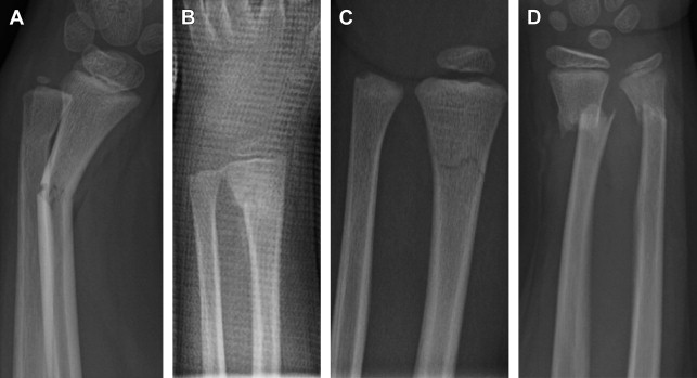
Metaphyseal Fractures
See Fig. 1 for 4 different examples of metaphyseal fracture patterns.
Torus fractures
A torus or buckle fracture refers to a unicortical, metaphyseal fracture most often resulting from a fall onto an outstretched hand. The cortex under compression, most commonly the dorsal cortex, fails or buckles, whereas the cortex under tension, most commonly the volar cortex, remains intact. Because of the intact cortex, these fractures are inherently stable. On examination, significant swelling or deformity is usually not seen. Point tenderness on the distal radial metaphysis confirms the diagnosis.
Torus fractures are treated with a short-arm splint or cast for 3 weeks, and radiographic follow-up of these injuries is typically not necessary. Studies have demonstrated that casting may not be necessary to ensure satisfactory healing. The application of a soft bandage or removable splint has been successfully used to treat these injuries.
Greenstick fracture
An incomplete fracture involving failure of the tension-sided cortex and plastic deformation of the compression cortex is termed a greenstick fracture. As described by Evans, this injury classically occurs as a result of a compression and rotational deformity. A distal third forearm greenstick fracture most commonly demonstrates an apex volar angulation and represents a supination injury. The reduction maneuver, aiming to counteract the deforming force, involves pronation of the forearm. Alternatively, an apex dorsally angulated fracture, representing a pronation injury, is reduced with supination of the forearm. Correction of the rotational deformity has been shown to be a reliable and easily reproducible reduction maneuver.
Bicortical Fractures
Nondisplaced fractures
Bicortical, or complete, fractures involving the distal radial metaphysis typically result from falls onto an outstretched hand but involve higher energy mechanisms than buckle fractures. Patients with these fractures frequently have associated distal ulna fractures, especially if torsion is combined with axial loading through the outstretched hand. Patients with nondisplaced, bicortical distal radius fractures typically present with pain and swelling about the wrist. On examination, the distal radius is tender to palpation on the metaphysis. For those with associated distal ulna fractures, the metaphysis, styloid, and the triangular fibrocartilage complex (TFCC) may also be painful and tender to touch. Active pronation/supination of the forearm and flexion/extension of the wrist are generally limited secondary to pain. Radiographs reveal a fracture line that extends transversely through the metaphysis.
A well-molded short-arm or-long arm cast is the recommended treatment of these nondisplaced metaphyseal fractures. In the author’s experience, patients with nondisplaced fractures of both the radius and ulna and those with painful forearm rotation are more comfortable in a long-arm cast initially. Radiographs should be obtained again at 7 to 10 days after injury to confirm that reduction has been maintained. The cast is removed at 4 to 6 weeks after injury. Adequate healing is confirmed by physical examination and repeat radiographs that show bone healing. After cast removal, instructions are given for range of motion and strengthening exercises; physical therapy is rarely needed. Within 8 to 10 weeks, patients may resume sports and other activities.
Displaced fractures
Patients with displaced fractures of the distal radius metaphysis typically present with a deformity of the wrist. Skin compromise at the fracture site, such as a small laceration or an abrasion with active bleeding, may indicate an open fracture. Neurovascular examination must be documented before reduction is attempted. Because most displaced fractures demonstrate dorsal displacement, the clinician should assess for volar wounds and median nerve injury. Sterile dressing of open wounds and provisional splinting should be done in the emergency department before obtaining radiographs to lessen the risk of ongoing soft-tissue injury and for patient comfort.
Closed reduction
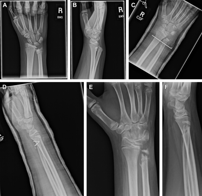
Displaced fractures are best treated with closed reduction and immobilization under conscious sedation in the emergency department. Fracture reduction may be facilitated by re-creation of the deformity that would relax the intact periosteum on the compression side of the fracture and allow the distal fracture fragment to slide over the proximal fragment. A well-molded sugar tong splint or cast would help maintain the reduction. When a cast is applied in the acute fracture setting, consideration should be given to bivalve the cast to accommodate subsequent swelling ( Fig. 2 ).
After reduction and application of a cast or splint, postreduction radiographs and a repeat clinical examination are mandatory. Most patients may be discharged home with fracture care instructions. Those patients with significant pain, severe swelling, abnormal findings on examination, or a potentially unsuitable home environment are best observed in the hospital overnight. On the hospital floor, instructions are given for strict wrist elevation, frequent neurovascular checks, and pain control that permits reliable evaluation but does not mask the signs of an evolving compartment syndrome or acute carpal tunnel syndrome.
Acute carpal tunnel syndrome, although rare in the pediatric population, has been reported after Salter-Harris (SH) 2 fractures of the distal radius. This complication is most common after dorsally angulated and displaced metaphyseal or physeal distal radius fractures in older children and adolescents. Patients developing acute carpal tunnel syndrome initially present with parasthesias in the sensory distribution of the median nerve. Differentiating a contusion to the median nerve from acute carpal tunnel syndrome can be challenging. The diagnosis is largely clinical and relies heavily on the progression of symptoms. A median nerve contusion presents as numbness or tingling in the volar aspect of the thumb, index, and long fingers that begins immediately after the injury. The symptoms are nonprogressive and tend to respond to elevation of the extremity and loosening of the dressings. Carpal tunnel syndrome, on the other hand, presents as a gradual progression of symptoms over a few hours after injury. The patient reports no relief with elevation and loosening of dressing. In the setting of progressive median nerve symptoms unrelieved by elevation, an urgent carpal tunnel release is indicated.
Acceptable Reduction of Metaphyseal Fractures
The distal radial physis accounts for 60% of the growth of the radius and typically closes at 14 to 16 years of age. A significant and predictable amount of remodeling occurs in fractures that heal with angulation and displacement if the physis is not injured. Acceptable reduction parameters vary with age ( Table 1 ).
| Age (y) | Sagittal Plane (°) | Coronal Plane (°) |
|---|---|---|
| 4–9 | 15–20 | 15 |
| 9–11 | 10–15 | 5 |
| 11–13 | 10 | 0 |
| >13 | 0–5 | 0 |
Owing to significant degree of remodeling, incomplete reduction of distal radial metaphyseal fractures may yield successful outcomes in children. Crawford and colleagues reported on 51 consecutive patients younger than 10 years with completely displaced distal radius fractures. The fracture was treated with gentle manipulation without sedation or local anesthetics to achieve angulation within 10° of normal. Translation and overriding of the fracture fragments (bayonet opposition) in the sagittal plane was accepted. At follow-up, all patients demonstrated union with full range of motion and radiographic evidence of remodeling. The achievement of a successful outcome without the potential complications and cost of conscious sedation certainly questions the traditional management of these fractures.
Cast considerations
Cast index
Several methods to evaluate casting have been developed, all with the purpose of quantifying the characteristics of casting technique that effectively maintains reduction. In the author’s opinion, and based on the published literature, the cast index, defined by the distance between inner cast edges measured on the lateral radiograph divided by that measured on the anteroposterior radiograph, is most easily calculated and a reasonable predictor of casting success. Chess and colleagues earlier had demonstrated the importance of the cast index in their study of 558 pediatric patients who underwent casting for forearm and wrist fractures. A significant difference in cast index was noted in those patients who had lost the initial fracture reduction. Webb and colleagues presented 113 patients with distal radius fractures treated with either a long- or short-arm cast. Patients who had failed to maintain the initial reduction had a significantly higher cast index, 0.79, than patients in whom the reduction was maintained, 0.71.
Short-arm versus long-arm casting
Controversy exists regarding the optimal length of the cast that best maintains reduction of displaced fractures. Traditionally, long-arm casts have been used after reduction of displaced distal radius. It was thought that this enhanced maintenance of reduction because elbow motion, more specifically pronation and supination, was restricted. Studies have shown, however, that short-arm casting may be equally efficacious in maintaining reduction. The quality of reduction and cast molding are more important factors in the prevention of late displacement than the length of the cast. Advocates of short-arm casting also demonstrated an advantage for patients, documenting that patients treated with below-elbow casts missed fewer days of school and required less assistance with activities of daily living than patients in long-arm casts.
Outpatient follow-up
Regardless of the length of cast or quality of reduction, late fracture displacement is common. Approximately a third of displaced distal radius fractures lose reduction, emphasizing the importance of close radiographic follow-up. Several factors may increase the risk of late displacement, including initial displacement (>50% translation, >30° angulation, bayonet apposition), incomplete reduction, concomitant distal ulna fracture, and poor casting technique. Weekly clinical and radiographic follow-up for 2 to 3 weeks after reduction is recommended to ensure that displacement is identified before fracture healing with malunion. Repeat closed fracture reduction of metaphyseal fractures may be safe for up to 2 to 3 weeks from injury. For most fractures, cast immobilization is used for a total of 4 to 6 weeks. Return to full activities can be expected at 2 to 3 months after injury, depending on the age of the child.
Physeal Fractures of the Distal Radius
Fractures of the distal radial physis are among the most common growth plate injuries seen in the pediatric population. Most of these injuries are SH 1 and 2 fractures ( Table 2 ). The risk of growth arrest after distal radius physeal fracture is about 4%. Although rare, it is the possibility of growth disturbance that sets these fractures apart from metaphyseal fractures. Although the short-term management of these fractures mirrors closely that of metaphyseal fractures, important differences must be noted.
| Salter-Harris 1 | 22% |
| Salter-Harris 2 | 58% |
| Salter-Harris 3 | 2.6% |
| Salter-Harris 4 | 2.0% |
| Salter-Harris 5 | 0.4% |
Nondisplaced fractures
Nondisplaced or minimally displaced fractures of the distal radial physis are common. The patient typically complains of wrist pain after a fall onto an outstretched hand without deformity or significant swelling. The examination is notable only for point tenderness on the distal radial physis. Radiographs may reveal a nondisplaced or minimally displaced fracture, most commonly SH 1 or 2 fractures. These fractures are treated with 3 to 4 weeks of immobilization similar to a torus or buckle fracture of the metaphysis. Often, however, radiographs show normal findings. In this scenario, it is the author’s practice to diagnose these injuries as occult SH 1 fractures. These injuries are often confused with wrist sprain or contusion. Owing to pain with activities and risk of a subsequent injury that can lead to displacement, these injuries are treated with cast immobilization.
Displaced fractures
Closed reduction
SH 1 and 2 fractures constitute the vast majority of displaced distal radius physeal fractures. Although closed reduction under conscious sedation in the emergency department is the best method of initial treatment, the surgeon must be careful to avoid excessive forceful or aggressive maneuvers for reduction, emphasizing instead the use of longitudinal traction and gentle repositioning to limit shear forces across the physis. Multiple attempts at reduction should also be avoided. Finally, because physeal healing is more rapid than healing of metaphyseal fractures, attempts at closed reduction of displaced SH 1 and 2 fractures later than 7 to 10 days after initial injury are not recommended. Not only is it likely that realignment will not be achieved, late reduction attempts increase the risk of iatrogenic physeal injury and growth arrest. Owing to the tremendous remodeling potential of these injuries, observation of deformity for remodeling is preferred over improving alignment at the risk of injuring the physis.
Owing to rapid healing of physeal fractures compared with metaphyseal fractures, repeat clinical examination and radiographs within 5 to 7 days of injury are recommended to ensure that loss of alignment is identified in such time that safe repeat closed reduction can be performed. Cast immobilization is maintained for around 4 weeks. By then, healing of distal radial physeal fractures is typically complete, as evidenced by resolution of point tenderness and radiographic healing. After cast removal, a removable splint may be applied and instructions for home exercises are given. Full return to activities can be expected within 8 to 12 weeks after injury. Because of the risk of growth arrest, patients should be followed up with radiographs at 6 monthly intervals till growth is documented or till skeletal maturity.
Salter-Harris 3 and 4 Fractures
SH 3 and 4 fractures are rare, occurring predominantly in older patients and in those with high-energy injuries associated with axial loading of the distal radius. At times, plain radiographs alone may not permit adequate assessment of the fracture pattern and displacement. In these cases, CT evaluation may be necessary. Nondisplaced fractures are treated similar to nondisplaced SH 1 and 2 fractures. Displaced fractures require special attention. Because these fractures are intraarticular and involve the physis, achieving anatomic or near-anatomic alignment is essential to improve the chances of successful outcomes. In some cases closed reduction and casting may achieve reduction, whereas most cases require percutaneous manipulation and pinning or open reduction and fixation.
Distal Ulna Fractures
Metaphyseal fractures
Distal ulnar metaphyseal fractures occur most commonly in the setting of an ipsilateral distal radius fracture after a fall from height onto an outstretched hand. Most distal ulna fractures achieve reduction indirectly during reduction of the distal radius fractures. Complete displacement with bayonet apposition and angulation of 20° to 30° are acceptable if the parameters of the radial reduction are within the accepted limits of remodeling. In rare cases, percutaneous reduction and pinning or open reduction are indicated for distal ulna fractures with unacceptable alignment. Healing of distal ulna metaphyseal is achieved in most cases by the cast immobilization used to treat the radius fracture.
Ulnar styloid and distal ulnar physeal fractures
SH fractures of the distal ulna are uncommon. SH 1 and 2 fractures most commonly occur in combination with distal radius fractures. As with metaphyseal ulna fractures, reduction of displaced fractures frequently occurs passively with reduction of the radius fracture and heal uneventfully. Clinical and radiographic follow-up of these fractures is important because of the risk of growth arrest resulting in ulnar shortening, a potentially problematic complication of these injuries. Growth arrest of the distal ulna has been reported to occur in 55% of physeal injuries, a figure dramatically higher than that seen in the distal radius.
In most cases, fractures of the ulnar styloid occur in the setting of fractures of the distal radius in children and adolescents. Cast immobilization used to treat the radius fractures leads to healing of some ulnar styloid fractures, but most do not unite radiographically. Despite nonunion, the vast majority of patients demonstrate excellent clinical outcomes without complaints of ulnar-sided wrist pain, instability, or functional limitation because in most cases, the TFCC remains intact and distal radioulnar joint (DRUJ) stability is not compromised. Symptomatic nonunion of the ulnar styloid presents with painful clicking and ulnar-sided wrist pain. This condition is best treated with excision of the nonunion and fixation of the TFCC to the base of the ulnar styloid. The rare fracture through the base of the ulnar styloid, often a result of high-energy trauma, is associated with DRUJ instability. When such a fracture is identified, some researchers recommend treatment with tension-band fixation and TFCC repair.
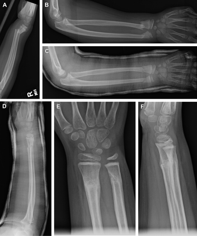
Pediatric Galeazzi fracture
Distal radius fractures with associated DRUJ disruption, commonly termed Galeazzi fractures, are most often the result of axial loading of the wrist with extreme pronation. In the pediatric patient, DRUJ instability is most commonly a result of a displaced distal ulnar physeal injury ( Fig. 3 ). Unlike in adults, the ligaments that stabilize the DRUJ remain intact and are attached to the ulnar epiphyseal fracture fragment. Because of this, the vast majority of Galeazzi fractures in children may be managed successfully with closed reduction and cast immobilization, with the forearm in supination. Surgical intervention is reserved for irreducible DRUJ disruption, which most commonly occurs secondary to extensor carpi ulnaris tendon or periosteal interposition. Potential long-term sequelae of these injuries include limitations of supination and pronation, ulnar nerve dysfunction, and persistent DRUJ instability.