Diagnostic Imaging Techniques
Robert Schneider
Helene Pavlov
Numerous diagnostic imaging techniques may be used to supplement history, physical examination, and laboratory tests in the evaluation of bone and joint disease. The choice of the imaging technique to be used and the sequence of usage of the techniques depend on the sensitivity and specificity of the technique for a particular problem; on the availability, cost, and risk involved; and on the experience in its use.
The goal is to answer the question raised by the clinician in the shortest time at the least cost and risk to the patient. A prior consultation with the radiologist and providing clinical information when ordering an imaging examination will help the radiologist and technologist to tailor their examination to the problem under investigation.
Radiography is usually the initial diagnostic imaging method in the evaluation of bone and joint pain because it is readily available and is of relatively low cost compared to other imaging methods. It provides excellent detail of bone anatomy and soft tissue calcification. Lucency, sclerosis, and periosteal reaction indicate bone abnormality. Cartilage, muscle, ligaments, tendons, and synovial fluid all appear with the same soft tissue density on radiography, which limits evaluation of abnormalities of these tissues. Cartilage destruction can be diagnosed if narrowing of the joint space is present (Fig. 7-1). Bone erosions can be seen around the joints. Determination of osteoporosis can be difficult because variations in technique can affect the apparent density on radiograms. Bony alignment can be determined and measured. Synovitis may be detected in the knee, elbow, and ankle because of the displacement of adjacent fat pads, but it cannot be reliably detected in the hip and shoulder. Other imaging methods, including magnetic resonance imaging (MRI), radionuclide scanning, and ultrasonography may pick up the bone and joint abnormality when radiograms are normal (Fig. 7-2).
Radiographic real-time imaging is done by fluoroscopy. C-arm fluoroscopes, which can be rotated and tilted, aid in localization during surgical procedures (e.g., internal fixation of fractures and osteotomies) and invasive radiologic procedures (e.g., myelography, nerve root, facet, and epidural spine injections, percutaneous needle biopsy, discography, joint aspiration and injection, and arthrography) (see Fig. 7-2C). Fluoroscopy with video recording can also be used for the evaluation of motion. Care must be taken to limit fluoroscopic time to avoid excessive radiation exposure.
Radionuclide scanning
Bone scanning with use of technetium 99m phosphate complexes has been used most frequently in the evaluation of metastatic disease to the skeleton and has largely replaced routine radiographic skeletal surveys for this purpose except for multiple myeloma. It is also used for the evaluation of benign bone disease because abnormalities that are not visible on radiograms may be detected. Bone scanning detects physiologic changes in the bone, as compared to the anatomic changes seen on radiograms. Increased uptake of radionuclide on a bone scan is caused mainly by increased osteoblastic activity associated with new bone formation and to a lesser degree by increased blood flow to bone. This can result from numerous causes, including infection, tumor, fractures, or synovitis. Although bone scanning is sensitive in detecting abnormalities of the bones and joints, it is
not specific. Three-phase bone scanning, which includes blood flow and blood pool scans, as well as static images taken 2 to 4 hours or more after injection of the radionuclide, should be ordered for the evaluation of localized bone or joint pain. The early phases show vascularity, which may be helpful in diagnosing synovitis, infection, and soft tissue abnormalities. Single-photon emission computed tomography (SPECT) may provide more detail and can be helpful in diagnosing stress or traumatic spondylolysis and in detecting photopenic areas in avascular necrosis. Radionuclide bone scanning is most useful for screening the entire skeleton to localize the site of abnormality and also for detecting stress fractures, osteoid osteomas, and for evaluating painful joint prostheses.
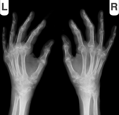
Figure 7-1. Oblique radiographs of the right and left hands and wrists in a woman with rheumatoid arthritis show narrowing of the radiocarpal, midcarpal, carpometacarpal, second metacarpophalangeal, and radioulnar joints, with periarticular osteoporosis and erosions.
Radionuclide infection scanning
Scanning with gallium citrate 67 shows increased uptake at sites of infection and some malignant neoplasms in the bones or soft tissues. It has a high sensitivity for bone and joint infection but is nonspecific because it may show increased uptake associated with other causes of increased bone turnover, including fractures or tumors, and may also show increased uptake in noninfectious inflammatory conditions, such as inflammatory arthritis. The specificity of a gallium scan for infection may be greater if it is compared with a bone scan. If the gallium scan shows more intense uptake than the bone scan at the affected site or if the uptake of gallium is not congruent with the uptake on the bone scan, then infection is likely. However, only one-third or even less of
bone infections meet these criteria. False-negative gallium scans may be seen in chronic infection or if the patient is treated with antibiotics before the scan is performed. Gallium scanning is preferred over white blood cell (WBC) scanning for diagnosis of infectious spondylitis, and for fever of unknown origin.Related posts:

Stay updated, free articles. Join our Telegram channel

Full access? Get Clinical Tree


