Diagnosis
Pauline M. Camacho
Osteoporosis is a skeletal disorder characterized by compromised bone strength, which predisposes to increased risk of fractures [1,2]. Bone strength is a product of both bone density and bone quality. Bone density is expressed as grams of mineral per area or volume; bone quality refers to factors such as architecture, turnover, damage accumulation (e.g., microfractures), and mineralization [2]. Whereas bone density can be measured by various methods that are clinically available, bone quality is not readily quantifiable.
Burden of the Disease
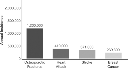
Contrary to other common diseases that produce distinct symptoms, osteoporosis can exist undetected for a long time before complications occur. It is estimated that this bone loss afflicts up to 28 million Americans; of these, 10 million have established osteoporosis [3]. Vertebral fractures are more common than breast cancer, stroke, and heart attack [3,4,5] (Fig. 1.1). A woman left untreated is predicted to have a 50% chance of suffering from an osteoporotic fracture sometime in her life [6].
As the cost of healthcare in the United States and other countries continues to rise, it is becoming evident that the complications arising from this preventable and treatable disease account for an increasing chunk of our healthcare spending. With a cost of approximately $21,000 per hip fracture, the estimated total cost of treating hip fractures worldwide in the year 2050 will be $131.5 billion [7]. Thus, it is important that physicians and patients take measures to prevent and treat the disease.
Clinical Picture
Height Loss
Even without measuring bone mass using available technology, certain symptoms and signs should clue the physician to the presence of the disease. Perhaps the most common, yet the least noted during clinic visits, is
height loss. The majority of vertebral fractures are asymptomatic, with the only presenting symptom being height loss and dorsal kyphosis. Recent findings from the risedronate pivotal trials have shown that height loss of more than 2 cm over 3 years has a sensitivity of 35.5% for detecting new vertebral fractures, and specificity was 93.6% [8]. Furthermore, one study showed that height loss is highly predictive of low spine and hip bone mineral density (BMD) [9].
height loss. The majority of vertebral fractures are asymptomatic, with the only presenting symptom being height loss and dorsal kyphosis. Recent findings from the risedronate pivotal trials have shown that height loss of more than 2 cm over 3 years has a sensitivity of 35.5% for detecting new vertebral fractures, and specificity was 93.6% [8]. Furthermore, one study showed that height loss is highly predictive of low spine and hip bone mineral density (BMD) [9].
 Figure 1.1. Osteoporotic fractures in women, compared with other diseases. Approximately 1.2 million osteoporosis-related fractures occur in women each year [3]. The incidence of osteoporosis-related fractures in women is greater than the annual combined incidence of heart attack [4], stroke [4], and breast cancer [5]. |
Dorsal Kyphosis
Dorsal kyphosis, or “dowager’s hump,” usually occurs as a result of multiple anterior wedge deformities in the thoracic and lumbar spine. It is important that clinicians explain to patients that this deformity is a result of osteoporotic fractures because even today, some patients still equate the “hump” with bad posture. Severe kyphosis also leads to impaired motion of respiratory muscles, leading to dyspnea and possibly restrictive lung disease.
Fragility Fractures
Fragility fractures are those that do not result from high trauma. These ostoeoporotic fractures usually result from falls. Other diseases that may cause low-trauma fractures include osteomalacia and metastatic bone disease.
In addition to vertebral fractures, common fracture locations include the wrist (Colles fractures), ankles, and hip, the fracture that carries the worst prognosis. Fractures in the pelvic bone do occur but are not typical of osteoporotic fractures. Occurrence of such should prompt an evaluation for osteomalacia or metastatic disease.
Acute compression fractures of the spine most commonly occur in the midthoracic region and present with sudden severe pain and tenderness. They usually occur while stooping or carrying a load, but sometimes occur with very little provocation. The pain usually requires multiple medications, such as analgesics and narcotics, for control and can last for weeks to months.
Patients with multiple compression fractures can suffer from chronic back pain, particularly in the lower back area. Hip fractures carry a mortality rate of up to 26%, most commonly due to associated complications such as infections and thromboembolic and cardiovascular events. Long-term disability and loss of independent function can occur in up to 50% of these patients [7,10,11,12].
Diagnosis
Osteoporosis is most commonly diagnosed using bone densitometry. Various techniques are available to quantify bone mass (Table 1.1), but the most accurate and precise is the central dual-energy x-ray absorptiometry (DXA) scan.
Basic Principle
The basic principle of the DXA scan is that a beam of x-ray is generated and is allowed to pass through the area of interest, usually the spine or the hip. The density of the bone, which is usually determined by its calcium content, causes varying degrees of attenuation of the x-ray beam. As the beam passes through bone and soft tissue, two photoelectric peaks are quantified, and the device is able to subtract the contribution of soft tissue to the measured density. BMD is expressed as an area measurement in grams per square centimeter.
Bone Density and Fracture Risk
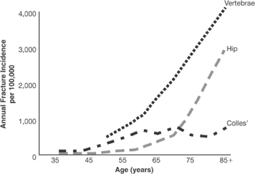
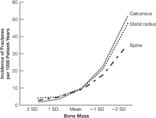
Fracture risk increases significantly with age (Fig. 1.2), and the incidence rises sharply after the menopausal years. Hip fractures occur about a decade later, with a sharp increase in incidence around age 70. A strong correlation exists between fracture risk and bone density, and it is said that this relationship is even stronger than that between cholesterol and heart disease (Fig. 1.3).
Table 1.1. Bone Measurement Techniques | |||||||||||||||||||||
|---|---|---|---|---|---|---|---|---|---|---|---|---|---|---|---|---|---|---|---|---|---|
| |||||||||||||||||||||
 Figure 1.2. Fracture risk with aging in white women. (Adapted from Riggs BL, Melton LJ III. Involutional osteoporosis. N Engl J Med 1986;314:1676, with permission.) |
 Figure 1.3. Fracture risk vs. bone density. There is an exponential relationship between decreasing bone mass and increasing incidence of fractures. (Adapted from Miller PD, Bonnick SL, Rosen CJ. Consensus of an international panel on the clinical utility of bone mass measurements in the detection of low bone mass in the adult population. Calcif Tissue Int 1996;58:207–214, with permission.)
Stay updated, free articles. Join our Telegram channel
Full access? Get Clinical Tree
 Get Clinical Tree app for offline access

|


