Crystal-Induced Arthritis
Joan M. Von Feldt
Gout
CLINICAL POINTS
Renal insufficiency is the most common cause of gout.
The condition is more common in men but is also found in postmenopausal women.
Monoarticular arthritis of the first metatarsal joint can be the first presenting sign.
Abrupt onset of pain, with inflammation, is characteristic.
INTRODUCTION
Gout is one of the oldest known types of arthritis. It was first identified by the Egyptians in 2640 BC, and podagra was later described by Hippocrates in the fifth century AD as the “unwalkable disease.” He noted the clinical correlation between podagra and its association with the wealthy. The word gout is derived from the Latin word “gutta,” which means a “drop” from the belief that gout was cause by drops of evil humors or, more specifically, drops of an excess of one of the four humors. This imbalance was believed to be the cause of gout. Among many of its sufferers are Benjamin Franklin, Thomas Jefferson, and Dr. Sydenham who in 1683 gave a classic description of an excruciating attack. Although the association between gout and uric acid has been known for centuries, it was only after the biochemistry of uric acid was elucidated in the 1960s that has led to better treatment and prevention of gout. Today, it is one of the most successfully treated forms of arthritis.
EPIDEMIOLOGY
Gout is predominantly a disease of adult men, with its peak incidence in the fifth decade. The prevalence of self-reported gout in the United States is estimated to be 3.9% of the US population (5.9% M; 2% F) and has been increasing in the United States over the past two decades, attributable to the increasing epidemic of obesity and concomitant hypertension.1
PATHOGENESIS OF HYPERURICEMIA
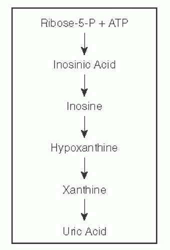
Uric acid is the normal end product of degradation of purine compounds. Its solubility in plasma is approximately 6.7 mg/dL at 37°C. Uric acid is derived from both ingestion of purines and the endogenous synthesis of purine nucleotides. Generation of uric acid involves the catabolic steps of degradation of hypoxanthine and xanthine. The major route of uric acid disposal is renal excretion, which accounts for approximately two-thirds of urate loss. See Figure 61-1 for a basic diagram of purine metabolism.
CLASSIFICATION OF GOUT
Both increased uric acid production and decreased uric acid renal secretion contribute substantially to hyperuricemia. The most common cause of gout is decreased uric acid excretion due to renal insufficiency. Traditionally, patients with gout have been subcategorized into two major categories: uric acid overproducers and underexcretors. To distinguish the two categories, 24-hour urine collections are calculated for uric acid concentrations. A person is classified as a uric acid overproducer if there is more than 750 mg of uric acid. If there is <750 mg of uric acid, then the patient is classified as an underexcretor. See Table 61-1 for the most common causes of overproduction/underexcretion and Table 61-3 for the most common causes of gout.
STAGES OF GOUT
The course of gout tends to pass through three distinct stages: asymptomatic hyperuricemia, acute intermittent gout (AIG), and chronic tophaceous gout (CTG) (Table 61-2). Asymptomatic hyperuricemia is defined physiologically as a level above the normal soluble concentration of monosodium urate crystals in body fluid, that is, 6.8 mg/dL. In women, the levels are lower but approximate the levels in men after menopause. The next stage starts with the initial attack of gout. The onset of a gouty attack includes warmth, swelling, erythema, and pain of the affected joint. The most commonly associated joint is the first metatarsal joint—inflammation of the first MTP is termed podagra. Systemic manifestations, such as low-grade fever, chills, malaise, and an elevated white blood cells (WBC) or erythrocyte sedimentation rate, (more common with polyarticular gout), can confuse the diagnosis as a possible infectious process. After resolution of an acute attack, there can be periods of quiescence with long intervals between attacks. Over time, these intervals can become shorter or more joints may be involved, ultimately developing into CTG. This usually develops after years of intermittent gout. The involved joints can become persistently uncomfortable and swollen, and very often, clinically evident tophi are detected. Common locations for tophi include fingers, ears, knees, olecranon bursa, and other pressure points.
TABLE 61-1 Causes of Uric Acid Overproduction/Underexcretion | ||||||||||||||||||||||||||||||||||||||||
|---|---|---|---|---|---|---|---|---|---|---|---|---|---|---|---|---|---|---|---|---|---|---|---|---|---|---|---|---|---|---|---|---|---|---|---|---|---|---|---|---|
| ||||||||||||||||||||||||||||||||||||||||
STUDIES (LABS, X-RAYS)
Laboratory
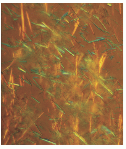
Elevated sUA is commonly seen in gout patients, but as reported earlier, asymptomatic hyperuricemia can precede true gout attacks by decades. In order to have a definitive diagnosis of gout, aspiration of synovial fluid and microscopy with polarized light showing bright, negatively birefringent needleshaped crystals must be performed. See Figure 61-2 for an example of monosodium urate crystals.
Radiology
Often, early in the course, there are no specific radiologic features of gout, except for possibly soft tissue swelling around the joint. However, in long-standing gout and, in particular, tophaceous gout, erosions may be present. These erosions are
usually distinguishable from other inflammatory arthritides in that a gouty erosion often has an “overhanging edge” with preserved joint space until very late in the disease.
usually distinguishable from other inflammatory arthritides in that a gouty erosion often has an “overhanging edge” with preserved joint space until very late in the disease.
TABLE 61-2 Stages of Gout | |||
|---|---|---|---|
|


