The olecranon process, coronoid process, and greater sigmoid notch are important components of the complex proximal ulna. Along with providing bony stability to the ulnohumeral joint, the proximal ulna serves as the attachment site of many important muscles and ligaments that impart soft tissue stability to the elbow joint. Management of proximal ulnar fractures continues to evolve as advances in imaging and anatomic and biomechanical studies have led to improvements in available implants; however, controversies remain, as shown in the current relevant literature.
Key points
- •
The olecranon and coronoid processes are important components of the complex proximal ulna, providing bony stability and attachment sites for many important muscles and ligaments.
- •
Most olecranon fractures are best treated operatively with tension-band wiring or plate fixation; plate fixation appears to be preferable for comminuted fractures.
- •
Some isolated displaced (>2 mm) olecranon fractures in elderly patients can be successfully treated nonoperatively.
- •
Coronoid fractures generally should be treated operatively, regardless of size, if there is associated instability; both the “lasso” technique and the suture anchors have produced good outcomes.
- •
Anteromedial facet fractures deserve special attention because of the facet’s contribution to elbow stability; most fractures require rigid fixation unless small and clinical examination demonstrates elbow stability.
Anatomic considerations
The anatomy of the proximal ulna is complex, and restoration of anatomic alignment is essential to restore normal biomechanics and avoid early arthritis, subluxation, and loss of function. Several studies have described anatomic features of the ulna that must be restored. Recently, much has been published on the proximal ulna dorsal angulation (PUDA) ( Fig. 1 ). Rouleau and colleagues characterized the PUDA in 50 patients examining bilateral elbow radiographs. They found a PUDA to be present in 96% of study participants with an average PUDA of 5.7° (range, 0–14). The average tip to apex distance was 47 mm (range, 34–78 mm). Both measurements were found to have good intrareliability and interreliability.
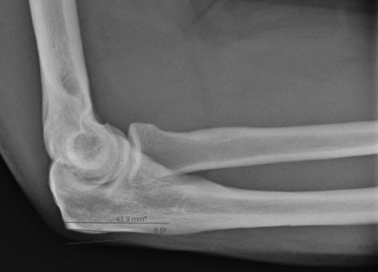
Imaging
Historically, imaging evaluation of proximal ulnar fractures has consisted of a standard anteroposterior and lateral elbow series supplemented with a radiocapitellar view as needed ( Fig. 2 ). However, recent improvements in imaging techniques and availability of 3-dimensional (3D) computed tomographic (CT) reconstructions have led some investigators to recommend more advanced imaging in certain fracture patterns. In their retrospective review evaluating the use of 2.7-mm and 2.4-mm plates, Wellman and colleagues found that 6 of 7 fractures thought to be a simple pattern on preoperative radiographs had occult comminution on CT scan. These investigators cautioned against using tension-band wiring (TBW) for fixation because many fractures may displace because of comminution not seen on standard plain films.
Olecranon fractures
Nonoperative Management
Olecranon fractures generally are managed operatively; however, nonoperative management is indicated in select cases. Conventional teaching has recommended that fractures with less than 2 mm of articular displacement and with an intact extensor mechanism should be treated nonoperatively. Duckworth and colleagues reported encouraging results with nonoperative treatment of isolated displaced (>2 mm) olecranon fractures in elderly patients. In their series of 43 patients with a mean age of 76 years, 72% had good to excellent results at 4 months, a Disabilities of the Arm, Shoulder, and Hand (DASH) score of 2.9, and an Oxford elbow score of 47, with a 91% satisfaction at 6 years. Veras del Monte and colleagues also reported good results after nonoperative treatment of displaced olecranon fractures in 12 elderly (mean age 82 years) patients. At final follow-up (average 15 months), no patient had any limitation in activity of daily living, and 8 of the 12 were asymptomatic with acceptable ranges of motion (ROMs). Clinical results were good in 8, fair in 3, and poor in one despite 9 fibrous nonunions. Eleven of the 12 patients graded their treatment as excellent.
Excision and Triceps Advancement
Olecranon excision with triceps advancement has been advocated as a viable treatment option in elderly patients with a comminuted fracture proximal to the coronoid with intact medial collateral ligament, distal radioulnar joint, and intraosseous membrane. The indications, technique for triceps attachment, and amount of olecranon that can be excised remain controversial. In the largest study to date, Gartsman and colleagues compared 53 isolated displaced olecranon fractures treated with primary excision to 54 treated with open reduction and internal fixation (ORIF). The groups had similar motion, function, pain, elbow stability, and incidence of posttraumatic arthritis; however, the complication rate was much higher in the ORIF group (23% compared with 4% in the excision group). In addition, biomechanical testing showed no difference in elbow extension between groups.
The amount of olecranon that can be removed before instability occurs has been debated. McKeever and Buck stated that as much as 80% could be excised without compromising elbow stability; however, Bell and colleagues performed serial resections and triceps advancements on 8 cadaver specimens and determined elbow stability after various amounts of olecranon resection. They noted instability to varus-valgus angulation and ulnohumeral rotation after resection of as little as 12.5%, with a progressive increase in instability up to 75%. Gross instability was noted after resection of 87.5%. Gartsman and colleagues found that one of their 54 patients treated with excision developed instability when 75% of the olecranon was excised. An and colleagues evaluated elbow stability after varying amounts of proximal ulnar resection and found a linear decrease in elbow constraint with increasing amounts of resection. They concluded that with resection of more than 50%, instability may occur; however, Inhofe and Howard reported that 11 of 12 patients had good or excellent results after excision of as much as 70%.
Tension Band Wiring or Plate Fixation
Before the advent of contoured proximal ulnar plates, displaced olecranon fractures were traditionally managed with TBW. This technique, in which tensile forces from the triceps are converted to compressive forces at the articular surface, has been advocated by the Arbeitsgemeinschaft für Osteosynthesefragen group. Long-standing debate exists regarding the method of TBW and indications for its use. In recent years, fixation using one-third tubular, dynamic compression, pelvic reconstruction, and, most recently, anatomically precontoured plate fixation has been advocated.
TBW is most often recommended for simple, transverse olecranon fractures without distal extension. The TBW construct consists of 2 longitudinal Kirschner wires and a wire placed in a figure-of-8 fashion through the dorsal cortex distal to the fracture and looped over the bent ends of the Kirschner wires proximally that are buried under the triceps ( Fig. 3 ). The proper location of the Kirschner wires has been recommended to be in the anterior cortex for bicortical fixation; however, concern over damage to neurovascular structures and penetration of the proximal radioulnar joint has led some investigators to advocate fixation in the distal or proximal ulnar canal. Huang and colleagues reviewed 78 displaced olecranon fractures treated with TBW fixation with 3 different Kirschner wire placement techniques: proximal ulnar canal, anterior ulnar cortex, and distal ulnar canal. They found proximal pin migration and elbow irritation when the wires were placed in the proximal ulnar canal and advocated placement in the distal canal to obtain adequate purchase and avoid the risk associated with anterior cortical penetration. In addition, in a review of 62 patients by Chalidis and colleagues, there was no difference in pin loosening or back-out whether or not the anterior cortex was engaged.
The use of TBW in comminuted fractures is debatable. Hume and Wiss compared fixation with a one-third tubular plate to fixation with TBW in 41 patients with displaced olecranon fractures. Plate fixation took on average 25 minutes longer, but the frequency of symptomatic hardware was much higher in the TBW group (42% vs 5%), as was loss of reduction resulting in significant articular step-off (>2 mm) (53% vs 5%). Clinical and radiographic results also were better in the group with plate fixation. Cadaver studies have shown plate fixation to be more stable than TBW in a comminuted fracture model. The recent advent of anatomic locked precontoured plates has led some investigators to recommend locked plate fixation for fractures with distal extension, dorsal comminution, or a high-energy mechanism. Lan and colleagues compared the outcomes in 10 patients treated with nonlocked plating to those in 14 patients treated with anatomic locked plating and found no difference in Mayo Elbow Performance Index (MEPI), ROM, and patient satisfaction. Schliemann and colleagues compared TBW to locking compression plate fixation in displaced, noncomminuted olecranon fractures. With 13 patients in each group, at an average follow-up of 43 months, the investigators found more good to excellent results based on mayo elbow performance score (92% vs 77%) and less frequent need for hardware removal (7 vs 12) in the plate group, but radiographic and clinical outcomes were similar. They concluded that given the higher overall cost of locked plate fixation, TBW was the treatment of choice for Mayo IIA olecranon fractures ( Fig. 4 ). However, given the small number of patients in the study, it is possible that it was underpowered to show a clinical difference. Moreover, no cost analysis was performed to examine the additional cost of hardware removal. The rate of hardware removal following TBW or plate fixation has varied considerably in the literature, ranging from 11% to 80% for TBW and 0% to 51% for plate fixation. Anderson and colleagues reported on the use of the Mayo Congruent Elbow Plate System for displaced olecranon fractures and found that only 3 of 32 patients required hardware removal.
Coronoid fractures
The coronoid is important to the ulnohumeral joint stability. Along with the radial head, it provides resistance to posterior displacement of the ulna on the distal humerus. In addition, the anteromedial facet provides stability against varus force. Along with its bony contribution to stability of the elbow, the coronoid also serves to stabilize the elbow joint through its insertion of the brachialis muscle, the anterior joint capsule, and the medial ulnar collateral ligament. Much has been published regarding the classification, diagnosis, and management of these often complex injuries, but there is still no clear consensus.
Anatomy/Classification
Classification of coronoid fractures has typically been based on size using the Regan and Morrey classification. Many investigators contend that the O’Driscoll classification, which is based on a fracture pattern that is associated with the overall pattern of injury, is more helpful to guide treatment. Doornberg and Ring examined 67 coronoid fractures and was able to confirm a strong association between O’Driscoll fracture type and mechanism of injury. The pattern of injury was classified as an olecranon fracture-dislocation (anterior or posterior), terrible triad, or varus posteromedial rotational instability pattern. They showed that olecranon fracture-dislocations were associated with large (>50%) coronoid fractures in 22 of 24 patients, whereas 31 of 32 terrible triad patterns had associated small (<50%) coronoid fractures. Of the 11 patients in the varus posteromedial instability group, all had fractures of the anteromedial facet. However, there has been little published on how to properly measure the coronoid height. Matzon and colleagues used 35 cadaver arms and a 3D digitizing system to define coronoid anatomy. Their results suggest that coronoid height is best defined by the trough of the trochlear notch and the slope change of the distal coronoid process ( Fig. 5 ).


