Abstract
Even though new prevention techniques have been developed and are being used during thoraco-abdominal aortic repairs, spinal cord infarction remains a severe and relatively frequent complication of aortic surgery. Infarctions in the territory of the anterior spinal artery are considered the most common. Different clinical pictures related to spinal cord transverse extension wounds are drawn up. In this paper, we present a case report of a subject having presented an isolated motor deficit of the lower limbs and a favorable prognosis, suggesting selective involvement of the anterior horns of the spinal cord subsequent to surgical repair of an aortic dissection. We wish to review the relevant anatomical, clinical and diagnostic characteristics along with current techniques of spinal cord ischemia prevention during and after surgery.
Résumé
L’ischémie médullaire reste une complication sévère et relativement fréquente de la chirurgie aortique malgré le développement des techniques de protection de la moelle épinière en peropératoire. Les infarctus dans le territoire de l’artère spinale antérieure sont les plus classiques. Suivant l’extension transverse de la lésion médullaire, différents tableaux sémiologiques sont décrits. Nous rapportons ici l’observation d’un patient ayant présenté un déficit moteur pur et de pronostic favorable, évoquant une atteinte isolée des cornes antérieures de la moelle dans les suites de la chirurgie d’une dissection aortique. Nous proposons une mise au point sur les caractéristiques anatomiques, sémiologiques, diagnostiques et sur les techniques de prévention des ischémies médullaires per- et post-chirurgicales.
1
English version
1.1
Introduction
Even though new prevention techniques have been developed and are being used during thoraco-abdominal aortic repairs, spinal cord infarction remains a common and frequent complication of aortic surgery.
Estimates of the prevalence of paraplegia through spinal cord infarction following treatment for aneurysm of the thoracic or abdominal aorta have been highly variable, ranging from 2.3 to 23% .
A prospective study involving 571 patients has estimated the occurrence of paraplegia or paraparesis subsequent to aortic surgery, whatever the causes, at 8.3% .
The nature and relative severity of the neurological damage depend on the height location of the infarction, its transversal extension and individual anatomical variations regarding vascularization of the spinal cord.
Onset of the complications may be acute, sub-acute or chronic, and the neurological deficit may arise immediately (secondarily to spinal cord hypoperfusion) or belatedly (as a result of the consequences of prolonged hypoxia) .
Different neurophysiological characteristics have been described according to the anatamoclinical form of the infarction: anterior spinal artery syndrome is the most clinically frequent. Its severity depends on the transversal extension of the infarction. At most the anterior two-thirds of the spinal cord are involved, with massive motor deficit below the lesion, constant vesico-sphincter disorders, and reduced sensitivity to pain and temperature below the wound . Cases of the Brown-Séquard syndrome, with a more favorable prognosis, have likewise been reported . At the minimum, the lesions may be limited to “grey matter” structures, particularly the anterior horns being most exposed to ischemia. These lesions are consequently responsible for predominantly proximal or distal hypotonic, areflexic, pauci- or plurisegmental motor deficits in the upper or lower limbs, without sensory disorder. Longitudinal extension of the infarction is determined by the quality of the anastomoses of the anterior spinal artery. In the lumbosacral area depending on the Adamkiewicz artery, damage to the conus medullaris and the cauda equina, which is the factor determining the degree of severity of the vesico-sphincter and sexual disorders, varies according to anastomotic compensation caused by the 5th anterior lumboradicular artery.
From an overall standpoint, prognosis for these types of infarction is severe. In a study of 36 patients , mortality during hospitalization was 22.2%; at the time of discharge 57.1% of the surviving patients were confined to a wheelchair, 25% walked with assistive device, and 17.9% walked normally. The most pejorative functional prognosis pertained to dorsolumbar infarction .
In this paper, we report on our observations of a patient having presented an isolated motor deficit of the lower limbs and benefiting from a favorable prognosis following surgical repair of an aortic dissection, and we wish to review the relevant anatomical, clinical and diagnostic characteristics along with prevention techniques of spinal cord ischemia during and after surgery.
1.2
Observation
Mr. C., 66 years of age, a retired cellar master, was hospitalized on 11/18/2008 in the physical medicine and rehabilitation (PMR) unit of the Bordeaux hospital for treatment of paraplegia that occurred subsequent to surgical repair of a type I aortic dissection.
Mr. C. presented several cardiovascular risk factors: android obesity with BMI higher than 30, high blood pressure, type 2 non insulin-dependent diabetes.
Initial chest pain on 10/27/2008 was rapidly followed by diagnosis of a type I aortic dissection with grade 2 to 3 aortic insufficiency associated with moderate pericardial effusion. A chest scan revealed the aortic dissection with an entranceway in the ascending aorta: it extended to the iliac arteries. The false lumen was permeable throughout the dissection without visualized thrombosis, particularly at the anastomosis of the intercostal arteries. The patient underwent emergency surgery the same day and benefited from replacement of the ascending aorta and the anterior portion of the aortic arch up to the brachiocephalic arterial trunk (BCAT) with conservation of the aortic valve. The operation was conducted under extracorporeal circulation with multisite cannulation involving cannulation of the femoral vessels and selective cerebral perfusion via the right axillary artery. The operation was conducted under hemodilution and moderate general hypothermia (28 °C).
Given the emergency context, monitoring of the motor and somatosensory evoked potentials (MEP and SEP) was not carried out during the operation.
Upon awakening, isolated motor paraparesis was noted at the upper lumbar level (ASIA motor score 70/100, level L1, AIS score C). This early neurological complication justified draining the cerebrospinal fluid with spinal cord “decompression” in mind.
Spinal cord MRI was carried out within 24 hours of the operation and considered as normal.
At the 72nd hour, aggravation was observed with the occurrence of subtotal paraplegia (ASIA motor score 60/100, level T12, AIS score C). The patient presented urinary and fecal retention, and neuroperineal tests showed sensory sparing and areflexia, even though contraction of the anal sphincter was still perceived.
Over the following 2 weeks the paraplegia regressed asymmetrically, while monoplegia of the lower right limb persisted. Spontaneous urination resumed on the 10th postoperative day without any post-micturition residue. Mr. C. was admitted to the PMR unit 3 weeks after surgical repair of the aortic dissection.
Clinical examination on admission to the PMR unit yielded the following findings:
- •
monoplegia of the lower right limb, which was flaccid, associated with overt quadriceps muscle atrophy. Initial functional muscle strength (MRC scale) was: hip flexor and extensor; 1/5, knee flexor and extensor; 2/5, foot extensor; 1/5, foot flexor; 1/5 (ASIA motor score 80/100, level L1); there remained some hypotonia in comparison with the other lower limb, which had fully recovered;
- •
absent right Achilles tendon reflex and clear diminution of the right rotulian reflex;
- •
wholly normal results in sensitivity tests at the level of the two lower limbs (ASIA “touch” score, 112/112; ASIA “pinprick” score, 112/112); there was no neuropathic pain;
- •
no vesico-sphincter disorder, normal neuroperineal testing results;
- •
cutaneoplantar reflexes in flexion.
Concerning the patient’s functional incapacity at the time of admission, the Barthel index yielded a score of 50/100.
Given a clinical picture suggesting isolated peripheral neuromuscular impairment, an electroneuromyogram was performed on December 9, 2008, that is to say 6 weeks after discovery of the neurological deficit:
- •
a needle detector test showed signs of diffuse and acute denervation at the two lower limbs level, but it clearly predominated on the right; at rest, there was considerable fibrillation activity along with numerous fasciculations; contraction activity was markedly limited;
- •
a stimulo-detection test revealed perfectly preserved sensory potential.
The test results finally provided arguments favoring the hypothesized anterior pluriradicular or anterior horn impairments. On the other hand, the normal sensory potential eliminated the hypothesis of lumbosacral plexopathy.
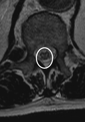
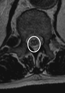
A second spinal cord MRI was carried out a month and a half after the surgery, and on the T2 sequences in axial sections it showed “hyperintensity predominating in the anterior grey matter structures and creating a ‘snake eyes’ aspect extending from T12 to L1”. As for the sagittal sections, a vertical linear hyperintensity was observed from T11 to L1 ( Figs. 1 and 2 ). These aspects were compatible with a spinal cord ischemia that could not be visualized on the first MRI performed in the immediate aftermath of the aortic surgery.


Diagnosis of a partial spinal cord infarction in the area of the Adamkiewicz artery was consequently validated, and a perioperative hypoperfusion mechanism was considered to be its likely cause.
Motor rehabilitation was carried on in a hospital setting for 2 months, and associated cardiovascular reconditioning under effort, gradual verticalization and gradual adapted muscle reinforcement of the knee extensors, and the hip extensors and abductors. Other forms of therapy included electromyostimulation sessions focusing on the damaged but recovering muscles, which were superimposed to voluntary contractions. The rehabilitation program also included walking resumption, initially with a system designed to lighten body weight, and then with the help of a long-leg orthosis. Lastly, the patient benefited from a program aimed at his regaining ambulatory autonomy thanks to assistive devices such as a walker and then crutches.
Neuromuscular assessment at the end of the hospital stay, that is to say three and a half months after the operation, confirmed pronounced improvement regarding the motor command: hip flexor, 3/5; hip extensor, 4/5; knee flexor, 4/5; knee extensor, 3/5, plantar flexor, 5/5; foot levators, 1/5 (ASIA motor score 92/100, AIS score D).
The rotulian and Achilles tendon reflexes remained diminished but were both evocable.
Notwithstanding the persistent weakness of the foot flexors and impaired “locking” of the extended knee, improved motor ability was functionally translated by efficient walking on crutches and use of a long-leg orthosis. Walking perimeter exceeded 150 meters on a flat surface.
Once discharged from the hospital, Mr. C. was able to return to his home and carry on his recovery with the assistance of a physiotherapist.
1.3
Discussion
Part 1: comprehension of the clinical syndrome of this patient justifies a preliminary summary of some anatomo-functional notions pertaining to vascularization of the spinal cord on the transversal and longitudinal planes.
Within the spine, there exist two arterial axes:
- •
the anterior axis is represented by the anterior spinal artery threading its way through the anterior median groove and governing vascularization of the anterior two-thirds of the spinal cord. Concerning the cervical spine, the anterior spinal artery stems either directly from the vertebral arteries or from the fusion of the radiculomedullary arteries, which themselves stem from the vertebral arteries. Concerning the dorsal spine, it stems from two or three intercostal arteries. Regarding the lumbosacral spine, anterior spinal vascularization is ensured by the Adamkiewicz artery, which originates in an iliac artery, and which usually suffices to provide vascularization for the lumbosacral spine. Around 15% of the time, however, an additional radicular artery is directed towards the conus medullaris and is connected at the “anastomotic arch” described by Lazorthes. Known as the artery of Desproges-Gotteron, this artery stems either from the hypogastric (internal iliac) artery or directly from a primitive iliac artery. Recent anatomical studies conducted with young subjects confirmed the existence of an anastomosis between the lumbosacral arteries and the “anastomotic arch” of the conus medullaris, but this type of connection does not necessarily entail effective compensation in the event of severe and prolonged ischemia in the area of the Adamkiewicz artery ;
- •
the posterior axis is composed of two small posterior spinal arteries threading their way along the dorsal columns. They vascularize not only the latter, but also the front portion of the posterior horns of the grey matter structures.
As we pointed out in the introduction, ischemic lesions essentially involve the territory of the anterior spinal artery governing the central territory, that is to say the majority of the grey matter structures. Concretely speaking, the anterior spinal artery may be said to give birth to the perforating sulcocommissural arteries, around 200 of which exist; they are particularly numerous at the lumbar and cervical enlargements level. The central territory constitutes the final territory of anterior spinal vascularization ( dernier pré ), and it includes grey matter structures (except for the front portions of the posterior horns) and the adjacent portion of the anterolateral columns and the dorsal columns.
The circumferential peripheral territory is tributary to the dense “piemerian” arterial network, which is fed by the anterior spinal artery and the two posterior spinal arteries, and it includes the superficial part of the anterolateral and the posterior columns, along with the front portions of the posterior horns.
Part 2: spinal cord infarction is more frequent in the anterior spinal cord territory; this may be explained by the fact that the radiculomedullary arteries in the anterior territory are less numerous and more irregularly distributed than those in the posterior territory.
Infarctions are also most frequently found to be seated at the level of the cervical and lumbar enlargements ; this is explained by the less sizable metabolic demand of the thoracic spinal cord and by the effectiveness at that level of the longitudinal anastomosis of the anterior spinal artery stemming from the overlying or underlying territories.
Confirmation in our patient of an infarction in the area of the Adamkiewicz artery, an infarctus limited to the central anterior territory depending on the sulcocommissural arteries and spanning the T12-L1 metameric segments, is congruent with the mode of onset (discovery of infarction on awakening from aortic surgery). The electroclinical picture is compatible with damage to the motor neurons of the anterior horn. The anastomosis underlying the damaged metameric segments are probably effective, even though this has not been demonstrated by selective angiography, and their likely effectiveness may explain at least the lifting of a diaschisis phenomenon on the conus medullaris, and the rapid regression of vesico-sphincter disorders. Finally, the infarction was limited to the anterior horns and spared the peripheral spinal cord territory; clinical examination never showed damage to the ascending or descending pathways (no sensory disorders in the paralyzed region, no spastic pyramidal syndrome; conversely, areflexia secondarily evolved to hyporeflexia). The isolated and circumscribed nature of the anterior horn lesion surely explained a favorable evolution translating the fact that massive destruction of the motor neurons on the metameric segments under consideration did not take place.
Partial spinal cord infarctions of the central territory of the anterior spinal artery that are limited to the grey matter of the anterior horns have been described from an anatomo-clinical standpoint and experimentally confirmed . Yet, they would appear to occur quite rarely. Sub-acute clinical pictures simulating an amyotrophic lateral sclerosis syndrome limited to the lower limbs have been reported in association with extrinsic compressions of the Adamkiewicz artery by discopathy at the thoracolumbar junction, and the compression has been confirmed by selective angiography of the lumbar enlargement artery .
Liblau et al. and Fernandez et al. have described two cases of infarction involving the central territory of the Adamkiewicz artery in all its height . In the case described by Liblau et al. , the infarction was bilateral, responsible for flaccid paraplegia, without sensory disorder or sphincter dysfunction. In the case described by Fernandez et al. , the infarction was unilateral and responsible for homolateral monoplegia at the site of the lesion; the hypothesis explaining the unilateral nature of the damage consisted in the doubling of the anterior spinal anterior axis, which received from each of its sides a radiculomedullary artery. In a series of 12 cases of patients having survived aortic surgery for aneurysms and traumatic dissections and who were diagnosed with ischemic myelopathy, Mathé et al. insisted on the importance of grey matter lesions that may be deduced from the anatomo-clinical syndrome: four patients in the series who had dorsolumbar lesions without sacral damage nonetheless suffered from permanent motor lesion spanning four to eight low lumbodorsal metameric segments.
In our case, the patient did not present any preoperative neurological disorder, and a preoperative scan confirmed the aortic dissection without thrombosis of the false lumen. As is often the case, the dissection was spiral and probably responsible for avulsion of the intercostal arteries, which represented a factor of poor perfusion. Association of extracorporeal circulation in hypothermia with multisite cannulation most likely failed to sufficiently protect spinal cord vascularization.
Part 3: what is the impact of intraoperative spinal cord protection techniques during surgical aortic repair?
Several spinal cord protection principles have been developed and are aimed at limiting risk of ischemia and/or reestablishing spinal cord circulation .
While some of the techniques are of proven effectiveness, it is difficult to distinguish their efficiency per se; more often than not, they are used in association with others:
- •
early reimplantation of the intercostal arteries is carried out only in surgery of the descending thoracic aorta, and it may be associated with significantly reduced risk of spinal cord ischemia . Multivariate analysis of a series including 337 patients having undergone surgery for aneurysm of the thoraco-abdominal aorta (types I, II and III alike) has demonstrated a significant reduction in neurological complications following early reimplantation of the intercostal arteries from T9 to T10 . In the case presented, reimplantation of the intercostal arteries could not be envisioned due to the extreme surgical complexity encountered during the aortic dissections. An initial surgical approach is addressed first to the anterior mediastinum (sternotomy) so as to reach the ascending aorta (avoidance of the entranceway) and to the aortic arch and not the thorax (thoracotomy) so as to reach the descending thoracic aorta and the intercostal arteries;
- •
extracorporeal circulation is conducted by means of multisite cannulation with atrio-venous discharge (right atrium) and two arterial cannulations (femoral and right axillary) facilitating expansion of the territories to perfuse. It should be pointed out that in aortic dissections, the circulating false lumen may provoke an avulsion of numerous visceral arteries, and that a simultaneous antegrade and retrograde perfusion (via the true and false lumens) will reduce the risk of organ malperfusion. In order to protect each and every organ, body temperature is lowered to 28 °C at the moment extracorporeal circulation is partially interrupted, and axillary perfusion alone is maintained as recourse, thereby enabling perfusion of the brain parenchyma. The aorta is opened and inspected; without renewed access to the aortic cross, vascular reconstruction (“open anastomosis” without aortic clamping), will stop at the foot of the brachiocephalic artery trunk but at times, according to the lesions that have been found, it will also involve the aortic cross. Ischemia time (by circulatory arrest) is of decisive importance concerning the occurrence of neurological complications. In the overall framework of aneurysms of the descending thoracic artery, several retrospective studies have shown that the occurrence of neurological complications is directly connected to aortic clamp time . In the series studied by Svensson et al. , paraplegia incidence came to 27% when the aortic clamping process lasted around 60 minutes, and to only 8% when it did not exceed half an hour. The series studied by Coselli included 1250 patients having undergone surgery for type I and type II aortic aneurysms, of whom 667 had benefited from extracorporeal circulation. Paraplegia incidence was significantly lower in this group (4.5%) than in a group not having had extracorporeal circulation (11.2%, P = 0.019). These results are particularly significant regarding type II aneurysms;
- •
the systemic hypothermia carried out on our patient is supposed to improve tolerance towards ischemia by diminishing demand for oxygen . Regional hypothermia of the spinal cord has likewise shown itself to be effective ;
- •
draining the cephalorachidian liquid (CRL) is another frequently used technique, but it is employed only in surgery for aneurysms of the descending thoracic aorta. Clamping of the thoracic aorta causes heightened CRL pressure through heightened intracranial pressure and through modified distribution of venous return. The pressure increase entails a diminution in spinal cord perfusion pressure, while CRL subtraction reduces the elevation of intrarachidian pressure and may consequently be aimed at improving spinal cord pressure; it brings perfusion pressure back towards the norm. Several studies have shown a significant decrease in ischemic paraplegia incidence when CRL is subtracted , particularly in association with intrathecal papaverine injection, which was not performed in our patient. In the series of d’Estera et al. , 654 cases were studied. Frequency of neurological complications came to only 3.3% in patients having benefited from CRL draining associated with distal reperfusion, as opposed to 8.4% when these techniques were not used. In a therapeutic trial, Coselli and Lemaire randomized 145 patients presenting type I or type II aortic aneurysm into two groups: a control group without intraoperative CRL draining, and a second group with CRL draining. Their study results showed an 80% reduction of postoperative complications in the second group (13% in the control group versus 2.6%; P = 0.03). In the general framework of aortic dissections, organization of prophylactic CRL draining might be advisable, especially for patients presenting signs of preoperative spinal cord damage (this was not the case with Mr. C.);
- •
maintenance of optimal hemodynamic stability during and immediately after surgery assumes an essential role in avoidance of any and all risk of low blood pressure that would entail diminished spinal cord perfusion.
Part 4: intraoperative recording of motor evoked potentials (MEP) and sensory evoked potentials (SEP) facilitates screening for the risk of neurological complications and may also help to orient the practice of the surgeon and the anesthetist (increased CRL draining, guidance in reimplanting the intercostal arteries). However, this type of recording during aortic surgery remains controversial, and is difficult to carry out in cases of extreme emergency.
A study carried out in 1998, by Safi and his team , assessed the interest of electrophysiological monitoring during aortic surgery; irreversible intraoperative modification of the evoked potentials is significantly associated with immediate occurrence of a neurological deficit (results not found in delayed deficits). The alterations of MEP and SEP show the same sensitivity, even though SEPs are easier to generate and record in the context of anesthesia/resuscitation. On the other hand, reversible modifications of the evoked potentials are not significant. Finally, normality of evoked potentials is of poor negative predictive value, and it may, in spite of everything, be associated with a neurological deficit.
Part 5: the importance of medical imaging (selective angiography, MRI).
The role of imaging in spinal cord ischemia is double; when highlighting the occlusion of the anterior radiculomedullary artery, selective angiography allows confirmation of a spinal cord ischemia diagnosis. In practice, spinal cord angiography is performed to preoperatively locate the origin of the Adamkiewicz artery, and thereby limit the risk of neurological complications. Once this artery is situated, it is possible to orient reimplantation of the intercostal arteries and to envision the use of substitute networks during the operation; the surgeon would thereby reduce the risk of spinal cord ischemia occurrence. However, the emergency entailed by a ruptured aneurysm of aortic dissection does not allow for selective angiography, which is reserved for more “settled” vascular interventions. In actuality – and emergency permitting –, selective angiography is customarily supplanted by non-invasive techniques: CT angiography and MRI angiography. Several studies have compared the respective effectiveness of the two techniques. The rate of Adamkiewicz artery detection ranges from 67 to 100% with MRI angiography, and from 68 to 90% with CT angiography . MRI angiography is used preferentially.
In practice, spinal MRI is the first examination to be carried out when a spinal cord lesion is suspected. Nevertheless, a normal MRI in the first hours, which is what was recorded for our patient, cannot formally preclude a diagnosis of spinal cord infarction.
The MRI is carried out with an injection of gadolinium on the axial and the sagittal planes in T1 and T2-weighted sequences. A duly constituted infarction is translated by a hypersignal in T2 and an isosignal or hyposignal in T1. The spinal cord may be augmented in size as a result of a vasogenic edema . The T2-weighted sequence is more sensitive, as it facilitates pinpointing of the exact level and extent of the lesion (sagittal sections), and enables recognition of the arterial territory involved (axial sections).
Infarcts of the anterior spinal cord arterial territory predominate in the central territory, at the level of the grey matter structures, thereby providing an aspect described in terms of “snake eyes” .
Sequences of diffusion may enhance the sensitivity of the MRI during the acute phase . The infarction is translated by a clear hypersignal with a lowered coefficient of diffusion.
1.4
Conclusion
Notwithstanding the development and utilization of numerous protective measures for the spinal cord, ischemia remains a classical complication of aortic surgery. Until now, the relevant physiopathological mechanisms have yet to be wholly identified. The case reported here underlines some frequently encountered difficulties in diagnosis, many of which have to do with the anatomical particularities. The case of our patient is relatively exceptional given the limitation of ischemic lesion to the anterior horn of the lumbar enlargement; what is more, the lesions are lacking in height, and the clinical evolution was rapidly favorable. This overall picture prompts us to underline how important it is to clearly indicate the anatomo-clinical syndrome of the infarction, which is an essential criteria of functional prognosis, and we also wish to insist on the assistance provided by imaging, especially spinal MRI, in our attempts to better understand the mechanisms of the lesion.
Disclosure of interest
The authors declare that they have no conflicts of interest concerning this article.
2
Version française
2.1
Introduction
L’ischémie médullaire reste une complication fréquente et classique de la chirurgie aortique malgré le développement des techniques de protection de la moelle épinière en peropératoire.
La prévalence d’une paraplégie par infarctus médullaire au décours d’une cure d’anévrisme de l’aorte thoracique ou abdominale est estimée de manière très variable entre 2,3 à 23 % .
Une étude prospective récente regroupant 571 patients, a estimé l’incidence de survenue d’une paraplégie ou paraparésie à 8,3 % au décours d’une chirurgie aortique toutes causes confondues .
La nature et la sévérité du déficit neurologique dépendent de la localisation en hauteur de l’infarctus, de son extension transversale et des variations anatomiques individuelles concernant la vascularisation médullaire.
Le mode d’installation peut-être aigu, subaigu ou chronique ; le déficit peut être immédiat (secondaire à l’hypoperfusion médullaire) ou retardé (en rapport avec les dommages de l’hypoxie prolongée) .
Différents tableaux séméiologiques ont été décrits selon la forme anatomoclinique de l’infarctus : le syndrome de l’artère spinale antérieure est la forme clinique la plus fréquente. Sa gravité est fonction de l’extension transversale de l’infarctus. Au maximum les deux tiers antérieurs de la moelle sont concernés avec un déficit moteur massif sous-lésionnel, des troubles vésicosphinctériens constants, un déficit sensitif sous-lésionnel à la douleur et à la température . Des tableaux de syndrome de Brown-Séquard, de meilleur pronostic, ont été rapportés . Au minimum, les lésions peuvent être limitées à la substance grise, notamment des cornes antérieures qui sont les plus exposées à l’ischémie. Ces lésions sont alors responsables de déficits moteurs hypotoniques, aréflexiques, pauci- ou plurisegmentaires à prédominance proximale ou distale aux membres supérieurs ou inférieurs, sans trouble sensitif. L’extension longitudinale de l’infarctus dépend de la qualité des anastomoses de l’artère spinale antérieure. Dans le territoire lombosacré dépendant de l’artère d’Adamkiewicz, l’atteinte du cône médullaire et de la queue de cheval, qui détermine la sévérité des troubles vésicosphinctériens et sexuels, est fonction de la compensation anastomotique apportée par la cinquième artère lombo-radiculaire antérieure.
Globalement, le pronostic de ces infarctus est sévère. Sur une étude de 36 cas , la mortalité pendant l’hospitalisation était de 22,2 % ; à la sortie 57,1 % des patients survivants étaient en fauteuil roulant, 25 % marchaient avec des aides techniques, 17,9 % avaient une marche normale. Le pronostic fonctionnel le plus péjoratif concernait les infarctus dorsolombaires .
Nous rapportons ici l’observation d’un patient ayant présenté un déficit moteur pur des membres inférieurs avec un pronostic favorable, dans les suites de la chirurgie d’une dissection aortique et proposons une mise au point sur les caractéristiques anatomiques, sémiologiques, diagnostiques et sur les techniques de prévention des ischémies médullaires per- et post-chirurgicales.
2.2
Observation
Monsieur C., 66 ans, maître de chais à la retraite, est hospitalisé le 18/11/2008 dans le service de médecine physique et de réadaptation (MPR) pour la prise en charge d’une paraplégie survenue dans les suites d’une dissection aortique de type I opérée.
Monsieur C. présentait plusieurs facteurs de risques cardiovasculaires : obésité androïde avec IMC supérieur à 30, hypertension artérielle, diabète de type 2 non insulinorequérant.
Une douleur thoracique inaugurale le 27/10/2008 a permis de porter rapidement le diagnostic d’une dissection aortique de type I avec insuffisance aortique de grade 2 à 3, associée à un épanchement péricardique modéré. Le scanner thoracique retrouvait la dissection aortique avec une porte d’entrée dans l’aorte ascendante et s’étendant jusqu’aux artères iliaques. Le faux chenal était perméable tout au long de la dissection sans thrombose visualisée notamment au niveau de l’abouchement des artères intercostales. Le patient a été opéré en urgence le même jour et a bénéficié d’un remplacement de l’aorte ascendante et de la partie antérieure de la crosse aortique jusqu’au tronc artériel brachiocéphalique (TABC) avec conservation de la valve aortique. La chirurgie a été menée sous circulation extracorporelle avec canulation multisite comprenant une canulation des vaisseaux fémoraux et une perfusion cérébrale sélective via l’artère axillaire droite. L’intervention était menée sous hémodilution et sous hypothermie générale modérée (28 °C).
Le monitorage des potentiels évoqués moteurs et somesthésiques (PEM, PES) n’a pas été effectué en peropératoire vu le contexte de l’urgence.
Au réveil, une paraparésie motrice pure de niveau lombaire supérieur était constatée (ASIA moteur 70/100, niveau L1, score AIS C). Cette complication neurologique précoce a justifié la mise en place d’un drainage du LCR dans la perspective d’une « décompression » médullaire.
L’IRM médullaire réalisée moins de 24 heures après l’intervention était considérée comme normale.
Une aggravation à la 72 e heure était constatée avec la survenue d’une paraplégie subtotale (ASIA moteur 60/100, niveau T12, score AIS C). Le patient présentait une rétention urinaire et fécale ; l’examen neuropérinéal montrait une épargne sensitive et une aréflexie, mais la contraction du sphincter anal restait perçue.
Dans les deux semaines suivantes, la paraplégie a régressé de façon asymétrique, laissant persister une monoplégie du membre inférieur droit. Les mictions spontanées ont été reprises au dixième jour postopératoire sans résidu post-mictionnel. L’admission de Monsieur C. dans le service de MPR est survenue trois semaines après la chirurgie de la dissection aortique.
L’examen clinique à l’entrée en MPR permettait de retenir les éléments suivants :
- •
une monoplégie du membre inférieur droit, flasque, associée à une amyotrophie quadricipitale franche. Les cotations musculaires initiales étaient respectivement : flexion/extension de hanche : 1/5, flexion/extension de genou : 2/5, flexion plantaire : 1/5, releveurs du pied : 1/5. (ASIA moteur 80/100, niveau L1) ; il persistait un certain degré d’hypotonie par rapport à l’autre membre inférieur qui avait totalement récupéré ;
- •
une abolition du réflexe achilléen droit et une nette diminution du réflexe rotulien droit ;
- •
une normalité de l’examen de la sensibilité, à tous les modes, au niveau des deux membres inférieurs. (ASIA sensitif « toucher » : 112/112, « piqûre » : 112/112). Il n’y avait pas de douleur neuropathique ;
- •
une absence de trouble vésicosphinctérien et un examen neuropérinéal normalisé ;
- •
des réflexes cutanéo-plantaires en flexion.
Concernant le niveau d’incapacité fonctionnelle du patient, l’index de Barthel à l’entrée était à 50/100.
Devant ce tableau évoquant une atteinte neuromusculaire motrice pure périphérique, un électroneuromyogramme a été réalisé le 09/12/2008, soit six semaines après la découverte du déficit neurologique :
- •
l’examen de détection à l’aiguille montrait des signes de dénervation aiguë diffuse au niveau des deux membres inférieurs, mais prédominant nettement à droite avec, au repos, une importante activité de fibrillation et de nombreuses fasciculations ; le tracé de contraction était très appauvri ;
- •
l’examen de stimulo-détection retrouvait des potentiels sensitifs parfaitement conservés.
Cet examen permettait finalement de retenir des arguments en faveur d’une atteinte soit pluriradiculaire antérieure, soit de la corne antérieure. L’hypothèse d’une plexopathie lombosacrée pouvait être écartée, en raison de la normalité des potentiels sensitifs.
Une seconde IRM médullaire a été réalisée un mois et demi après la chirurgie. Celle-ci montrait, sur les coupes axiales pondérées en T2, une « hyperintensité prédominant dans la substance grise antérieure, réalisant un aspect en « yeux de serpent », étendue de T12 à L1 » . Sur les coupes sagittales on notait un hypersignal linéaire vertical de T11 à L1 ( Fig. 1 et 2 ). Ces aspects étaient compatibles avec une ischémie médullaire, non visualisable sur la première IRM effectuée dans les suites immédiates de la chirurgie aortique.


