This article discusses the diagnostic criteria, clinical course, and complications of complex regional pain syndrome. Multidisciplinary treatment including physical and occupational therapy, psychological evaluation and treatment, pharmacologic management, and more aggressive options including sympathetic blocks, sympathectomy, and spinal cord stimulation are also reviewed.
Key points
- •
Complex regional pain syndrome (CRPS) is characterized by pain out of proportion to the usual time or degree of a specific lesion.
- •
The diagnosis of CRPS is based on 4 distinct subgroups of signs and symptoms: sensory, vasomotor, sudomotor, and motor/trophic changes.
- •
Treatment should be multidisciplinary, consisting of medications, physical/occupational therapy, psychotherapy, and sympathetic blocks targeted toward pain relief and functional restoration.
- •
More aggressive treatment, such as sympathectomy and spinal cord stimulation, have a low level of evidence but may be considered for therapy-resistant CRPS type I.
Introduction
Complex regional pain syndrome (CRPS) is characterized by pain that is out of proportion to the usual time or degree of a specific lesion. It does not present within the distribution of one peripheral nerve or nerve root, and has a distal predominance of abnormal sensory, motor, sudomotor, vasomotor, and/or trophic findings. Progression is variable. CRPS has been known by many other names including reflex sympathetic dystrophy (RSD) and causalgia. These terms date back to Claude Bernard, who in 1851 referred to a pain syndrome that was accompanied by changes in the sympathetic nervous system. During the American Civil War, Silas Weir Mitchell described cases of soldiers suffering from ongoing burning pain after recovering from gunshot wounds, and coined the term Causalgia. Evans first used the term reflex sympathetic dystrophy in the 1940s to emphasize that the sympathetic nervous system was involved in the pathophysiology of the disease. CRPS replaced the term RSD for several reasons. Sympathetic changes and dystrophy may not be present throughout the disease course. Furthermore, there is no specific reflex arc that is responsible for the CRPS; pain is secondary to multisynaptic pathologic changes involving the brain, spinal cord, and peripheral nerves.
Epidemiology
CRPS has a female to male ratio of 2:1 to 4:1, which is more common with increasing age. There are 50,000 new cases of CRPS in the United States annually. The most common initiating events of the syndrome include fractures, sprains, and trauma such as crush injuries and surgery. Immobilization after injury is a contributing factor in more than half of patients.
Diagnosis
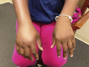
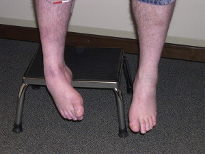
The Budapest Consensus Workshop introduced criteria to identify patients with CRPS and exclude other neuropathic conditions. More stringent criteria are used for research purposes to eliminate false-positive inclusions. Less stringent criteria are used in the clinical setting to avoid missing the diagnosis. A patient must report symptoms of, and display signs on physical examination, in the following categories: sensory, vasomotor, sudomotor/edema, and motor/trophic ( Figs. 1 and 2 ). For both clinical and research purposes, a patient with CRPS should have physical examination evidence of at least 1 sign in 2 or more of the categories. The symptom criteria are different when assessing patients in a clinical rather than a research setting. In a clinical setting, patients must report 1 symptom in 3 out of the 4 categories, whereas in the research setting the patient must report 1 symptom in each of the 4 categories ( Box 1 ). This minor adjustment in data collection creates a sensitivity of 0.85 and a specificity of 0.69 for the research group, compared with the clinical criteria that have a sensitivity of 0.94 and a specificity of 0.36.


- 1.
Continuing pain that is disproportionate to any inciting event
- 2.
Patient must report 1 symptom in 3 of the 4 following categories:
- a.
Sensory: Reports of hyperesthesia and/or allodynia
- b.
Vasomotor: Reports of temperature asymmetry and/or skin color changes and/or skin color asymmetry
- c.
Sudomotor/edema: Reports of edema and/or sweating changes and/or sweating asymmetry
- d.
Motor/trophic: Reports of decreased range of motion and/or motor dysfunction (weakness, tremor, dystonia) and/or trophic changes (hair, nail, and/or skin)
- a.
- 3.
Patient must have 1 sign at the time of evaluation in 2 or more of the following categories:
- a.
Sensory: Evidence of hyperalgesia to pin prick and/or allodynia to light touch and/or deep somatic pressure and/or joint movement
- b.
Vasomotor: Evidence of temperature asymmetry and/or skin color changes and/or asymmetry
- c.
Sudomotor/edema: Evidence of edema and/or sweating changes and/or sweating asymmetry
- d.
Motor/trophic: Evidence of decreased range of motion and/or motor dysfunction (weakness, tremor, dystonia) and/or trophic changes (hair, nail and/or skin)
- a.
- 4.
There is no other diagnosis that better explains the signs and symptoms
There are 2 subgroupings of CRPS. CRPS I is CRPS without major nerve damage (formerly known as RSD) while CRPS II is CRPS with major nerve damage (formerly known as causalgia). A third subtype is CRPS NOS (not otherwise specified), which captures patients who only partially meet the current criteria but were diagnosed with CRPS under previous criteria.
Accurate diagnosis of CRPS is challenging despite the standardization of diagnostic criteria. There is no one definitive objective test that confirms the clinical diagnosis. Physical findings may not be present at all times, but the diagnostic criteria for signs require that the findings be present at the time of the diagnosis. At present, the diagnosis is made primarily on the basis of physical examination, but there are objective tests that may help verify the physical examination findings. Functional imaging, visual analog scales, and devices to quantify temperature and mechanical allodynia are available. Vasomotor findings are supported with a thermometer and Doppler measurement of vasomotor tone. Edema is quantitated with volumetry. Sudomotor function can be measured directly with quantitative sudomotor axon response testing, and indirectly with biopedance and skin potential fluctuations. Weakness and range of motion are measured by clinicians. Bone density testing is available. Small-fiber dropout via skin biopsy can be measured to validate a decrease in small nerve density. Most of these technologies are not readily available in the office.
Diagnostic testing may include rheumatologic workup to assess for inflammatory arthritis. Electrodiagnostic testing serves to evaluate the peripheral nervous system, which can help in the diagnosis of CRPS II. Plain films may reveal advanced osteoporosis or fracture in the symptomatic limb with CRPS. Magnetic resonance imaging evaluates soft-tissue injuries and bone edema. A triple-phase bone scan is generally not diagnostic of CRPS.
Pain may or may not be mediated by the sympathetic nervous system. A fluoroscopic guided lumbar paravertebral block for the lower extremity and a stellate ganglion block for the upper extremity may be useful in determining how much sympathetic input contributes to a patient’s symptoms. Because CRPS may or may not have a sympathetic component, a positive or negative response to a sympathetic block does not substantiate the diagnosis of CRPS.
Progression and course of disease
Schwartzman and colleagues report that after 1 year most of the signs and symptoms are well developed. Most patients have abnormalities of pain processing such as allodynia, which is present in 90% of patients at 5 years and in 98% of patients by 15 years. Swelling is noted in 75% of patients at 5 years and 90% by 15 years. Loss of strength and difficult movement is seen in 90% of patients at 5 years. Spread of the pain occurs in 92% of patients.
In a study of 27 patients with CRPS I, Maleki and colleagues reported contiguous spread in all patients, 70% of whom had independent spread to another site, 15% mirror-image spread to the initial site, and 19% contiguous spread alone. Van Rijn and colleagues reported that CRPS usually affects one limb but can spread to the contralateral or ipsilateral limb in 53% and 30% of cases, respectively. A diagonal spread was seen the least in 14% of cases. Aberrant regulation of neurogenic inflammation, maladaptive neuroplasticity, and genetic predisposition are theorized as the pathophysiology behind the spread of CRPS. Spontaneous spread is at the level of the spinal cord, as opposed to a systemic etiology.
The CRPS Severity Score (CSS) was developed in an attempt to assess the severity of CRPS. The concept of staging CRPS has been abandoned owing to the lack of empirical statistical evidence to suggest the existence of the stages. The CSS is based on the presence or absence of 17 clinically assessed signs and symptoms. Patients with higher CSS scores had greater reported pain intensity, distress, and functional impairments. Greater temperature asymmetry and abnormalities in thermal perception were seen more frequently.
Psychiatric issues
There is a debate as to whether patients who have CRPS are predisposed to develop the condition based on their psychiatric profile. Harden and colleagues found that preoperative anxiety and severity of pain predicted the development of CRPS signs and symptoms following total knee arthroplasty. However, most studies do not find a unique relationship between CRPS and psychiatric factors. Shiri and colleagues compared psychological profiles of patients with CRPS with those of patients suffering from conversion disorders. High somatization and depression and low anxiety scores were seen in both groups. Reedijk and colleagues compared patients with CRPS I–related dystonia with those with conversion disorders and affective disorders. Although the CRPS patients did exhibit elevated scores for somatoform dissociation, traumatic experiences, general psychopathology, and lower quality of life compared with the general population, they also had lower total scores for personality traits, recent life events, and general psychopathology relative to the patients with conversion disorder and affective disorder. The investigators concluded that patients with CRPS I–related dystonia did not have a uniquely disturbed psychological profile as a group. Puchalski and Zyluk reported that 62 patients who underwent distal radius fractures and developed CRPS did not exhibit significant differences in personality or depression scales relative to patients who did not develop CRPS. Monti and colleagues compared 25 CRPS I patients with a control group with chronic back pain. Both groups exhibited similar findings of major depressive and personality disorders. It was concluded that the abnormal findings are a result of severe chronic pain and are not uniquely secondary to CRPS.
Beerthuizen and colleagues conducted a systematic review of the literature since 1980, and concluded that there was no relationship between psychological factors and CRPS; they concluded that CRPS was associated only with patients who experienced more life events (divorce, death of spouse, vacation, and so forth). Geertzen and colleagues also reported that a difficult time in life or a painful affective loss (stressful life event) is more common during the onset of CRPS when a group of patients with subacute CRPS were compared with a group of patients preparing for hand surgery over the next 24 hours.
Treatment
In 1997, consensus guidelines were generated for the functional restoration of CRPS. Medication, psychological counseling, and interventional options were reserved for patients who were failing to progress with physical and occupational therapy. Current literature supports the use of medication, modalities, interventions, and psychological treatment more acutely. Multiple articles support the use of physical therapy to treat CRPS. Interdisciplinary treatment of CRPS is supported without high-level evidence by the aforementioned consensus-building conferences. Interdisciplinary treatment of the more general category of chronic pain has been used for decades. The objectives of physical and occupational therapy in patients with CRPS are to minimize edema, desensitize a painful limb and normalize sensation, promote normal positioning, decrease muscle guarding, and increase functional use of the extremity. Edema management consists of specialized compressive garments along with manual edema mobilization techniques. Aquatic therapy has also been shown to aid in edema control and to facilitate early weight bearing.
Desensitization can be achieved through a stress-loading program consisting of scrubbing and carrying techniques. Scrubbing involves using the affected limb to move a brush against a surface. Carrying involves a gradual weight-loading program whereby the patient carries objects and weights either in the hand or in a handled bag. For the lower extremities, weight-shifting and balancing techniques are used to gradually stress the affected leg. Contrast baths can also be beneficial in mild cases of CRPS. Desensitization and improved circulation may be accomplished in the affected extremity by alternating vasodilation (heat) with vasoconstriction (cold).
Patient education involves explaining fear-avoidance models using the symptoms, beliefs, and behaviors of the individual patients. Patients are taught to view their various autonomic and vasomotor disturbances as a condition that can be self-managed, rather than a disease whereby the affected limb needs careful protection. Under therapist supervision, the patient identifies dangerous and threatening situations and gradually increases exposure to these activities as much as possible until anxiety levels have decreased.
Mirror therapy, or mirror visual feedback, is conducted with the patient seated before a mirror that is oriented parallel to the midline. View of the affected limb is blocked behind the mirror. The patient first closes his or her eyes and describes both the affected and unaffected limb, followed by imagined movements of both extremities. When looking into the mirror, the patient sees the reflection of the unaffected limb positioned as the affected limb. The patient is then asked to look at the mirrored limb without movement. Finally, the patient moves the unaffected extremity through different planes of movement. Movement of or touch to the intact limb may be perceived as affecting the painful limb.
Graded motor imagery (GMI) acts on the reorganization of cortical networks presumed to be involved in chronic pain and CRPS. GMI consists of a sequential set of brain exercises comprising laterality training, imagined hand movements, and mirror feedback therapy. The patient looks at a photograph of a hand or foot, then imagines moving the painful limb into the position in the photograph. This action is progressed to moving both limbs into the position while observing the unaffected limb in a mirror that obscures the affected limb.
Alternative therapeutic techniques include acupuncture, Qigong therapy, and relaxation training. However, there is very low-quality evidence that any of these methods are effective in reducing CRPS I–related pain. Hyperbaric oxygen therapy has been reported to reduce pain and edema in a placebo-controlled, randomized study of 71 CRPS patients.
Stay updated, free articles. Join our Telegram channel

Full access? Get Clinical Tree


