1
English version
1.1
Introduction
Congenital orthopaedic disorders are not infrequent. Even though some orthopaedic disorders are immediately obvious at birth (such as congenital talipes equinovarus [CTEV]), others are not apparent or may even be hidden (such as congenital bone hypoplasia). This means that a comprehensive orthopaedic examination is of great value when screening for congenital orthopaedic disorders . Congenital talipes equinovarus is one of the most frequent congenital orthopaedic disorders. It may occur alone or in combination with other malformations. However, an association with hypoplasia of the navicular bone has never previously been described. Here, we report on a case of unilateral hypoplasia of the navicular bone discovered during the treatment of CTEV.
1.2
Case report
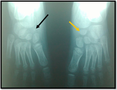
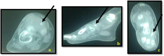
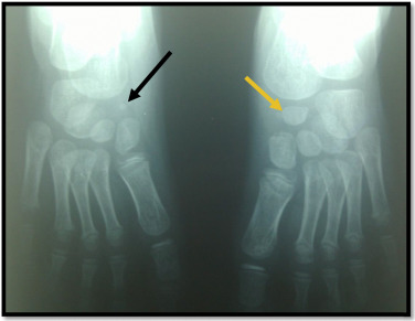
A 5-year-old child had been treated in another institution for right-side CTEV (initially with the Ponseti manipulation/plaster cast method, followed by use of a brace and orthopaedic insoles) since the age of 10 days. The condition had only improved slightly, prompting the patient’s family to consult in our clinic. On admission, a clinical examination revealed right-side CTEV with 30° of varus, 20° of supination and a 15° equinus deformity. The child had a Pirani score of 4 out of 6 and a Dimeglio score of 10 out of 20 . Standard X-rays of the feet were taken with a view to accounting for the absence of improvement – especially we did not have access to any previous radiological assessments. Anteroposterior and lateral X-rays of the feet ( Fig. 1 ) revealed hypoplasia of the navicular bone on the right side and the presence of a normal navicular bone on the left. We completed our assessment by performing a computed tomography scan of the right foot; the results confirmed the presence of hypoplasia of the navicular bone and the absence of any other bone abnormalities ( Fig. 2 ).


The child continued treatment with the Ponseti method, which consisted in the weekly application of plaster casts to progressively correct the forefoot over a 6-week period, followed by percutaneous tenotomy of the Achilles tendon and a 3-week cast immobilisation . As soon as the cast was removed, appropriate rehabilitative and proprioceptive work was initiated with a view to increasing joint range of motion, strengthening the lateral peroneus muscles and improving gait. The degree of deformity was significantly reduced, with a Pirani score of 0.5 out of 6 and a Demiglio score of 5 out of 20. However, the patient still had 15° of varus deformity, requiring the prescription of three-point insole.
1.3
Discussion
Congenital talipes equinovarus is one of the most frequent and most serious malformations of the foot . It may be combined with other malformations, which worsen the prognosis and emphasize the value of systematic screening. Several anatomical abnormalities may be combined with CTEV to various extents (such as agenesis or hypoplasia of the anterior or posterior tibial artery and fibular hemimelia).
Although abnormal features of the navicular bone have been described in patients with CTEV (e.g. a wedge-like shape or medial subluxation), the present report is the first to describe hypoplasia.
Hypoplasia of the navicular bone has never been observed before. After birth, foci of ossification appear at the cuboid bone, the three cuneiform bones and the navicular bone. The latter ossifies during the fourth year of life . A punctiform aspect at the age of five years (as was the case for our patient) is suggestive of hypoplasia, especially when the contralateral navicular bone is clearly visible and has a normal shape on a standard X-ray.
The value of radiographic assessment in the first months of life is debatable, and depends on the shape and the presence or absence of bony nodules. Some physicians perform radiological examinations on a regular basis, whereas others only do so in the event of a lack of correction (as was the case for our patient).
This case report emphasizes the importance of systematic screening for hidden abnormalities (such as hypoplasia) in cases of CTEV – especially when correctly performed treatment does not result in an improvement.
Disclosure of interest
The authors declare that they have no conflicts of interest concerning this article.
2
Version française
2.1
Introduction
Les déformations orthopédiques congénitales ne sont pas rares. Si certains troubles orthopédiques sont évidents dès la première observation du nouveau-né, comme le pied bot varus équin (PBVE), d’autres sont inapparents, voire cachés comme l’hypoplasie osseuse congénitale : d’où l’intérêt d’un examen orthopédique très complet dans une optique de dépistage . Le PBVE représente l’une des plus fréquentes malformations congénitales. Il peut être isolé ou associé à d’autres malformations. Son association avec l’hypoplasie de l’os naviculaire n’a jamais été décrite. Nous rapportant un cas d’hypoplasie unilatérale d’un os naviculaire découvert lors du suivi et de la prise en charge d’un PBVE.
2.2
Observation
Il s’agit d’un enfant de 5 ans aux antécédents de PBVE droit pris en charge initialement dans une autre institution depuis l’âge de 10 jours par des plâtres successifs selon la technique de Ponseti suivis d’attelle puis de semelles orthopédiques. L’évolution a été marquée par l’apparition d’une amélioration partielle ce qui a conduit la famille à nous consulter. L’examen à l’admission révélait un PBVE droit associant un varus de 30°, une supination de 20° et un équin de 15°. L’enfant avait un score de 4/6 selon Pirani et 10/20 selon Diméglio . Des radiographies standard des deux pieds ont été demandées dans le but d’expliquer l’absence d’amélioration d’autant plus que nous ne disposons pas de bilan radiologique antérieur. La radiographie des deux pieds de face et de profil ( Fig. 1 ) a révélé une hypoplasie de l’os naviculaire à droite, l’os naviculaire gauche était présent. Nous avons complété l’exploration par un scanner du pied droit qui a confirmé l’hypoplasie de l’os naviculaire sans autres anomalies osseuses associées ( Fig. 2 ).


