Clinical Diagnosis and Imaging
Brian D. Cameron
INTRODUCTION
Sources of Pain
Shoulder pain is a relatively common complaint that clinicians are asked to evaluate. The potential for successful treatment of shoulder pain is predicated on making the correct clinical diagnosis. Establishing the correct diagnosis or diagnoses can be particularly difficult. The clinician is faced with the challenge of identifying the source of pain and its clinical effect in anatomic and mechanical terms. Lack of an identifiable mechanical problem most likely will render an unreliable result. Although the clinical hallmark of glenohumeral arthritis is usually pain, arthritis is not the typical source of shoulder pain and dysfunction. Glenohumeral arthritis also may exist with other pathologic sources of pain from the periarticular soft tissues, associated joints of the shoulder girdle, or cervical spine (1, 2, 3, 4). The shoulder girdle is comprised of three diarthrodial joints (glenohumeral, acromioclavicular, sternoclavicular), two bursal articulations (scapulothoracic, acromiohumeral), four major muscle groups (scapulothoracic, thoracohumeral, scapulohumeral, and claviculohumeral), and four sets of ligaments (glenohumeral, acromioclavicular, coracoclavicular, and sternoclavicular). The causes of shoulder pain may be attributed to more than one of these structures. Pain related to nerve root, brachial plexus, or peripheral nerve lesions also may mimic primary shoulder pathology. The ensuing presentation of the painful shoulder therefore may be complex.
Glenohumeral Arthritis
Each of the glenohumeral arthritides is accompanied by varying degrees of secondary involvement of the synovium, capsule, glenohumeral ligaments, and rotator cuff. From the onset of the disease, the patient’s function deteriorates. Articular destruction is accompanied by gradual dysfunction of the soft tissues, leading to painful restriction of motion and functional impairment. As the disease progresses, the clinical manifestations resulting from articular erosion, ligamentous attenuation (or contracture), and rotator cuff dysfunction are often profound. These factors will influence the type of surgical intervention, choice of prosthetic reconstruction, and surgical outcome. Clinical evaluation requires a thorough clinical interview, skilled physical examination, and the judicious use of diagnostic tests. Successful diagnosis and treatment also require that the clinician have a thorough understanding of the glenohumeral arthritides and associated disorders. The main categories of glenohumeral arthritis include primary and secondary degenerative osteoarthritis, inflammatory arthritis, avascular necrosis (AVN), capsulorrhaphy arthropathy, and rotator cuff tear arthropathy. They may be further characterized as monarticular or polyarticular and acute or chronic. Once the disease process has been defined, the clinician is faced with the challenge of determining the appropriate course of management for each patient. Patients with glenohumeral arthritis often have a combination of mechanical, inflammatory, and psychologic components to their pain (5). The success of treatment is defined by its ability to ameliorate pain and restore function and a sense of well-being.
Treatment
In the presence of glenohumeral arthritis, pain is the primary indication for shoulder arthroplasty (6, 7, 8). Secondary indications include functional impairment and loss of motion. The diagnosis of arthritis is not in and of itself an indication for treatment. Rather, treatment should be considered only when the patient has perceived that the condition is affecting his or her quality of life. Nonsurgical treatment usually is initiated once the diagnosis has been established (9). This includes getting relative amounts of rest, avoiding provocative activities, and taking oral or intraarticular antiinflammatory medications (1). Shoulder mechanics may be preserved by using gentle, patient-conducted, range-of-motion and conditioning exercises (8). Once the disease has become recalcitrant to conservative measures, surgical intervention is considered. Non-prosthetic options include synovectomy, arthroscopic debridement of loose cartilage and osteophytes, open or arthroscopic capsular release, glenoidplasty, periarticular osteotomy, corrective osteotomy, resection arthroplasty, interpositional arthroplasty, and arthrodesis. Prosthetic options include hemiarthroplasty alone, hemiarthroplasty with interpositional glenoid resurfacing, and unconstrained or constrained total shoulder arthroplasty.
This chapter will address the major causes of glenohumeral arthritis. It also will highlight the clinical features and radiologic findings of these conditions and their influence on achieving a successful shoulder arthroplasty.
HISTORY AND ETIOLOGY
Patient Profile
A thorough history begins with an assessment of the primary complaint, usually pain. The location of arthritic pain is often vague, radiating into the anterior and posterior aspects of the joint line or into the axilla. The pain usually is exacerbated with recumbency; patients may experience inability to sleep on the affected side or pain that awakens them (1,6,10). Active and passive positioning of the arm will reproduce pain and frequently produce crepitance. Patients also may complain of restricted motion, weakness, atrophy, and tenderness over the anterior or posterior aspects of the joint line (10). The examiner also should investigate other common sources of pain, including rotator cuff, acromioclavicular joint, or neurogenic pathology. Pain in the contralateral shoulder, multiple joints, or cervical spine should be noted.
Acuteness of onset and duration of symptoms will vary depending on the arthritic condition. Previous trauma, fracture, dislocation, instability, infection, and surgery often are predisposing factors of arthritis. A complete medical and surgical history will assist in elucidating inflammatory, steroid-induced, and postsurgical etiologies. Patients should be asked specifically about any metabolic or rheumatologic conditions. Information regarding age, activity level, social history, patient motivation, and psychologic profile should be gathered. All of these factors will have a bearing on the treatment plan and will influence preoperative planning and patient counseling.
Age and General Health
No chronologic age limit exists, either young or old, for prosthetic shoulder arthroplasty (6). However, caution should be exercised when considering shoulder replacement for osteoarthritic patients who are young or place high functional demands on the upper extremity (paraplegia, heavy manual laborer, weight lifter). These patients possess a higher theoretic risk of mechanical glenoid component loosening and polyethylene wear-related aseptic loosening of the components following total shoulder arthroplasty (11). Hemiarthroplasty alone results in a high rate of progressive glenoid wear (12). Therefore, temporizing measures should be exhausted prior to shoulder arthroplasty in these patients. Patients whose occupational, recreational, or athletic activities place high physical demands on the shoulder may need to be counseled to avoid or delay the procedure or to consider a lifestyle alteration
that is commensurate with shoulder arthroplasty. The exception is the young patient with rheumatoid arthritis. Early total shoulder arthroplasty is preferred in patients with painful rheumatoid arthritis, good bone stock, and a competent rotator cuff (13). Most of these patients have polyarticular disease that limits their activity level and mitigates the risk of mechanical loosening of the shoulder arthroplasty.
that is commensurate with shoulder arthroplasty. The exception is the young patient with rheumatoid arthritis. Early total shoulder arthroplasty is preferred in patients with painful rheumatoid arthritis, good bone stock, and a competent rotator cuff (13). Most of these patients have polyarticular disease that limits their activity level and mitigates the risk of mechanical loosening of the shoulder arthroplasty.
Active elderly patients with painful shoulder arthritis usually will benefit significantly from shoulder arthroplasty (14, 15, 16). Medical and social issues that need to be addressed preoperatively include cardiac and anesthetic risks, the presence of dementia, and social support in the perioperative period. The elderly patient who is unable to mentally or physically comply with the postoperative rehabilitation program will obtain a poor result. The patient who normally requires the use of a walker for ambulation will require significant assistance for several months or will need to be counseled away from surgery.
General medical considerations include cessation of smoking, identifying and treating local or remote infections, and addressing dental problems. Cardiac and anesthetic risks should be assessed, and medications should be reviewed. Blood-thinning agents may need to be adjusted or temporarily discontinued prior to surgery.
Motivation
Patient compliance in the postoperative rehabilitation program is essential for a successful result (6,8). Inability or unwillingness to cooperate (dementia, psychiatric illness, drug or alcohol addiction, too busy) should be considered at least a temporary contraindication for surgery. Unrealistic expectations should be addressed prior to advocating prosthetic replacement. Expectations in activity level should be commensurate with the goal of longevity of the prosthesis. The patient also should have a thorough understanding of the risks, benefits, and limitations of the procedure.
Osteoarthritis
Osteoarthritis (degenerative joint disease) is recognized as the most common cause of musculoskeletal pain and dysfunction (1,17). The prevalence of glenohumeral osteoarthritis is less than that of the hip or knee and has the highest average age of onset (17,18). Although the cause of the disease is obscure, susceptibility may include genetic and ethnic predisposition, age association, and biomechanical factors (9). There is a higher incidence in elderly individuals, especially women (18,19). Younger individuals may develop osteoarthritis as a result of either acute or repetitive trauma. Glenoid dysplasia or hypoplasia may increase the risk of osteoarthritis (20). Static posterior subluxation of the humeral head may represent a previously unrecognized entity leading to osteoarthritis in the young adult (21). Glenohumeral osteoarthritis may be defined as either primary or secondary. Primary osteoarthritis typically presents without antecedent trauma. Secondary osteoarthritis may be related to chronic dislocations or instability or may be the result of surgical attempts to correct glenohumeral instability. Although the etiologies may differ, they share common pathologic features of progressive, irreversible destruction. The disease is mechanically driven and biochemically mediated. Whether the result of age-related inability to accommodate normal forces or failure to respond to excess loading, the process is an imbalance of reparative and degradative forces (17). It is characterized by asymmetric joint space narrowing, subchondral sclerosis, cyst formation, and the development of large osteophytes. Although the articular cartilage is most obviously affected, osteoarthritis is a disorder of the entire joint, including the bone, synovium, capsule, and glenohumeral ligaments.
Mechanical factors certainly play a role in the development of osteoarthritis, although the threshold for irreversible cartilage damage is less clear. Normal physiologic joint reactive forces result from muscle contraction and approach body weight as the arm is elevated between 60 and 90 degrees of abduction (9,22). Physiologic translational motion (shear force) is greatest with passive positioning of the shoulder and does not seem to result in permanent cartilage damage (23,24). Alterations in the magnitude, direction, or duration of loading may lead to adverse effects.
Secondary Osteoarthritis
Secondary, or posttraumatic, osteoarthritis generally results from an identifiable traumatic insult such as fracture, instability, or surgery. Alterations in anatomy, biology, or mechanics of the joint may lead to the development of arthritis.
Fracture
Intraarticular fractures resulting in articular incongruity will alter the normal distribution of contact forces across the joint. Head-splitting fractures are usually the result of high-velocity trauma in the young patient or occur in elderly patients. Marginal fractures of the glenoid may be associated with glenohumeral dislocations. Iatrogenic glenoid fractures also may occur as a complication of posterior glenoid osteotomy for posterior instability (26). The ability of the articular surface to tolerate changes in the joint contour depends on the surface area, articular geometry, loading characteristics, and stabilizing forces of the
ligaments and muscles. The glenoid normally covers 60% of the humeral head in the coronal plane and 46% of the head in the axial plane, resulting in a glenoid face that articulates with 28% of the humeral articular surface area (27). The tolerance of the glenoid to withstand excessive loading is probably less than that of the humeral head because the glenoid is relatively smaller and is experiencing constant loading by varying portions of the humeral head. A glenoid articular offset will decrease a surface contact area that is already small and will significantly increase the contact forces.
ligaments and muscles. The glenoid normally covers 60% of the humeral head in the coronal plane and 46% of the head in the axial plane, resulting in a glenoid face that articulates with 28% of the humeral articular surface area (27). The tolerance of the glenoid to withstand excessive loading is probably less than that of the humeral head because the glenoid is relatively smaller and is experiencing constant loading by varying portions of the humeral head. A glenoid articular offset will decrease a surface contact area that is already small and will significantly increase the contact forces.
Knowledge of the vascular anatomy may be helpful in predicting the risk of AVN following three- and four-part proximal humerus fractures. The reported rate of AVN associated with these complex fractures varies according to the severity of the injury, personality of the fracture, and method of treatment. Four-part fracture dislocations have an associated AVN rate approaching 100%, whereas valgus-impacted four-part fractures have an AVN rate of about 25% (28,29). Osteoarthritis will occur in more than half of the displaced four-part fractures. Three-part fractures have a lower rate of AVN (0% to 14%) and osteoarthritis (25%) (30). The risk of AVN is higher for open reduction and internal fixation of both three-part and four-part fractures as compared to closed (or limited open) reduction and minimal osteosynthesis (31, 32, 33, 34, 35). The clinical significance of osteonecrosis is mitigated by anatomic restoration of the fracture (36). Osteoarthritis in the setting of fracture may result from a combination of intraarticular injury, malunion, instability, vascular disruption, and capsular contracture (Fig. 6-1).
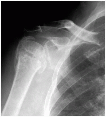 Figure 6-1 Osteonecrosis and osteoarthritis develop following a displaced three-part proximal humerus fracture. |
Dislocation Arthropathy
Dislocation arthropathy is an infrequent cause of arthritis and may be related to acute or recurrent instability, chronic dislocation, or iatrogenic sources. The index dislocation may occur in association with a fracture of the humeral head or glenoid, capsulolabral disruption, or rotator cuff tear. This may result in recurrent episodes of instability and repetitive chondral damage (37). However, there is no clear relationship between the presence (or severity) of arthropathy and number of dislocations (38). Neither is there a correlation with those patients treated surgically or nonoperatively (39). Arthritis may develop after successful surgery for the treatment of glenohumeral instability, even when normal shoulder motion and stability have been restored. Following dislocation, the incidence of developing some degree of resulting arthropathy is greater than 20%, and appears to be related to the primary dislocation (38). The risk of developing arthrosis that requires prosthetic arthroplasty is 10- to 20-fold greater in patients with a history of dislocation (40). Posterior dislocations hold a higher risk of developing arthritis, presumably because of a delay in diagnosis and treatment of the condition (39). It is interesting that some patients continue to demonstrate instability in the face of advanced arthritis (41).
Capsulorrhaphy Arthropathy
The term “capsulorrhaphy arthropathy” is used to describe the development of glenohumeral arthritis following surgical repair for the treatment of instability (6,7). Although this sequela may occur following treatment of any direction of instability, the common denominator is excessive tightening on one side of the joint (42). This complication occurs most commonly following a nonanatomic repair, most notably a Putti-Platt reconstruction (43, 44, 45, 46, 47, 48). It also may occur following a unidirectional repair in the treatment of multidirectional instability or excessive tightening of an “anatomic” repair (41). Excessive tension following an anterior repair will push the humeral head posteriorly and also will restrict external rotation (48). Attempts to externally rotate the arm will lead to further obligate posterior translation of the head relative to the glenoid. Eccentric loading leads to posterior glenoid erosion and rapid deterioration of the joint.
Inflammatory Arthritis
Inflammatory arthropathies are systemic illnesses with a genetic, autoimmune, or infectious association but no clear primary etiology (49,50). Common disorders include rheumatoid arthritis, ankylosing spondylitis, systemic lupus erythematous, psoriatic arthritis, gout, and pseudogout. Laboratory tests including serum chemistries, complete blood count, sedimentation rate, C-reactive protein, uric acid levels, and specialized screens (antinuclear antibodies [ANA] screens, rheumatoid factor [RF], human
leukocyte antigen [HLA]-B27) may be helpful in establishing or confirming the diagnosis, although this usually has been established prior to orthopedic evaluation (1). Aspiration of the shoulder should be considered in the presence of an acutely painful shoulder effusion. Attention should be directed at obtaining a cell count, gram stain, culture, and crystal analysis.
leukocyte antigen [HLA]-B27) may be helpful in establishing or confirming the diagnosis, although this usually has been established prior to orthopedic evaluation (1). Aspiration of the shoulder should be considered in the presence of an acutely painful shoulder effusion. Attention should be directed at obtaining a cell count, gram stain, culture, and crystal analysis.
Rheumatoid arthritis is the most common of these disorders and is representative of the pathologic and clinical manifestations seen at the glenohumeral joint. The ensuing discussion therefore will be directed primarily toward the symptomatic rheumatoid shoulder.
Shoulder pain occurs in 60% to 90% of patients with rheumatoid arthritis, with variable involvement of the acromioclavicular joint, subacromial bursa, rotator cuff, and glenohumeral joint (51,52). The clinical pattern is frequently one of symmetric, bilateral, polyarticular involvement. Although the shoulder is commonly affected, rarely is it the primary manifestation of the disease or the only involved joint. Isolated, acute, monarticular shoulder involvement should alert the clinician to the possibility of a septic joint (53,54). The clinical course of rheumatoid arthritis is more typically one of insidious onset and gradual progression of pain and dysfunction (1). Morning stiffness is common. Patients with rheumatoid arthritis often present notably late in the course of the disease with painful synovitis and swelling, severe arthritis, functional loss, and limitation of motion (6). Shoulder pain, especially nocturnal pain, is the most common complaint in patients with rheumatoid arthritis of the glenohumeral joint. However, the source of shoulder pain in the patient with rheumatoid arthritis is often difficult to isolate. Cervical radiculopathy and myelopathy frequently develop in patients with rheumatoid arthritis, leading to pain and weakness in the shoulder and upper extremity (49). Pain in the acromioclavicular joint, subacromial bursa, or rotator cuff may mimic (or coexist with) glenohumeral arthritis (1,10,50). Ipsilateral involvement of the elbow or wrist may be referred to the shoulder. A careful history and physical examination should be combined with radiographs and selective local anesthetic injections to identify the correct sources of pain and determine the appropriate treatment (55). Failure to address concomitant pathology at the time of shoulder arthroplasty may result in a poor outcome (52,56).
Cuff-Tear Arthropathy
Neer and colleagues first coined the term “cuff-tear arthropathy” in 1977, describing the uncommon sequela of untreated full-thickness rotator cuff tears (57). The normal rotator cuff centers the humeral head within the glenoid concavity. A balanced anterior and posterior force couple will center the head within the glenoid fossa, even in the presence of a full-thickness rotator cuff tear. In the absence of a functional rotator cuff, the humeral joint reaction force is not centered in the glenoid fossa as the arm is elevated. Instead, the unopposed pull of the deltoid directs the humeral head toward the superior margin of the glenoid. Narrowing of the acromiohumeral interval is a characteristic feature of the disorder (10,57,58). Erosions occur initially along the undersurface of the acromion and the superior surface of the humeral head, followed by the glenohumeral articulation (59). The conforming articulation that begins to develop from humeral head contact along the acromion, superior glenoid, and base of the coracoid sometimes is called “acetabularization.” The coracoacromial arch provides a new fulcrum for the humeral head and represents the final restraint to anterosuperior escape. Fluid may herniate through the acromioclavicular joint (57), resulting in the so-called “geyser sign” on arthrogram (60). Further rotator cuff degeneration accelerates articular wear and failure of the subchondral bone. Particulate joint debris accumulates and calcium phosphate crystals may be detected on analysis of the joint fluid (57,61). The end result is severe joint destruction in the presence of a massive, irreparable rotator cuff tear.
Cuff-tear arthropathy is seen more commonly in older individuals and females, is frequently bilateral, and may follow failed attempts at rotator cuff repair (10,57,62, 63, 64). Symptoms of pain and weakness generally have been present for years, punctuated by occasional painful exacerbations. Characteristically, the patient complains of unrelenting pain that radiates toward the deltoid tuberosity and is intensified with recumbency (10,57). Atrophy, swelling, crepitance, and functional deficits are also common complaints. Treatment prior to arthroplasty may include rotator cuff conditioning exercises, pain medication, intraarticular steroid injections, attempted rotator cuff repair, acromioplasty, rotator cuff debridement, and distal clavicle excision. Surgical intervention that compromises the integrity of the coracoacromial ligament will negatively affect the results of arthroplasty and often results in anterosuperior escape of the humeral head (62,64, 65, 66).
Related posts:


Stay updated, free articles. Join our Telegram channel

Full access? Get Clinical Tree


