Clinical Cases
Pauline M. Camacho
Paul D. Miller
Case 1
History
A 61-year-old white woman had a dual-energy x-ray absorptiometry (DXA) scan ordered by her primary-care physician. Other than mild essential hypertension, she had no other known medical problems. She fractured her wrist after a fall 3 years earlier. She had lost 2 inches in height since her 30s. After the DXA results were given to her, she started taking Viactiv calcium chews 3 times a day. Her mother may have suffered from osteoporosis. She denied a history of kidney stones, smoking, or excessive alcohol intake.
Physical Examination
A grade 2 thoracic kyphosis was detected. The rest of the examination was unremarkable.
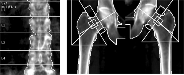
DXA Images
 |
| Region | BMD (g/cm2) | Young-Adult T-Score | Age-Matched Z-Score |
|---|---|---|---|
| L1 | 0.767 | -3.0 | -1.5 |
| L2 | 0.767 | -3.6 | -2.0 |
| L3 | 0.874 | -2.7 | -1.1 |
| L4 | 0.821 | -3.2 | -1.6 |
| L1-L2 | 0.767 | -3.3 | -1.8 |
| L1-L3 | 0.807 | -3.0 | -1.5 |
| L1-L4 | 0.811 | -3.1 | -1.5 |
| L2-L3 | 0.825 | -3.1 | -1.6 |
| L2-L4 | 0.823 | -3.1 | -1.6 |
| L3-L4 | 0.848 | -2.9 | 1.4 |
| Region | BMD (g/cm2) | Young-Adult T-Score | Age-Matched Z-Score |
|---|---|---|---|
| Neck | |||
| Left | 0.619 | -3.5 | -1.8 |
| Right | 0.647 | -3.3 | -1.6 |
| Mean | 0.633 | -3.4 | -1.7 |
| Difference | 0.027 | -0.2 | 0.2 |
| Laboratory Findings | |
| Hemoglobin (12–15 mg/dL) | 13 mg/dL |
| Serum creatinine (0.7–1.5 mg/dL) | 0.8 mg/dL |
| ALT (7–35 IU/L) | 23 IU/Ll |
| 25-OHD (target >30 ng/mL) | 24 ng/mL |
| Intact PTH (10–65 pg/mL) | 72 pg/mL |
| Ionized calcium (1.10–1.30 mmol/L) | 1.15 mmol/L |
| Total serum calcium (8.9–10.3 mg/dL) | 9.0 mg/dL |
| Phosphorus | 3.8 mg/dL |
| 24-hour urine calcium (100–300 mg/24 h) | 135 mg/24 h |
| Bone-specific alkaline phosphatase Premenopausal female: 4.5–16.9 μg/L Postmenopausal female: 7.0–22.4 μg/L Male 25 years and older: 6.5–20.1 μg/L | 21 μg/L |
| ALT, alanine aminotransferase; PTH, parathyroid hormone. | |
Teaching Points
Dr. Camacho
Patients frequently underestimate the significance of low trauma fractures. It is important to stress that fractures that result from a fall from a standing position are technically “fragility or osteoporotic fractures.”
This patient’s DXA image shows that she is osteoporotic by World Health Organization (WHO) criteria. Lumbar spine T-score is -3.1, and femoral neck T-score is -3.5. However, looking closely at the Z-scores, they fall between -1.6 and -2.0.
This implies that the patient is close to two standard deviations below other women her age in terms of bone density, highly suggesting premature bone loss.
A careful work-up for secondary causes of osteoporosis was warranted and subsequently revealed vitamin D deficiency and secondary hyperparathyroidism. Urinary calcium excretion is in the low-normal range.
Vitamin D deficiency should be treated, in this case with vitamin D 50,000 IU once a week for 3 months, subsequently reduced to 50,000 IU once a month. Various treatment regimens are discussed in Chapter 7.
Although vitamin D deficiency is frequently a result of poor nutritional intake and inadequate sun exposure in the elderly, the patient should be questioned about symptoms of malabsorption and a family history of celiac sprue. If clinically suggested, celiac antibodies must be obtained.
It has not yet been studied whether an antiresorptive agent should be given to such patients with vitamin D deficiency as part of the initial therapy. Although rare, there are reports of hypocalcemia induced by bisphosphonates in this population. This patient’s vitamin D levels were corrected to a current goal of greater than 30 ng/mL. One year after correction of her PTH levels to normal range, an antiresorptive agent was started.
Case 2
History
A 49-year-old white woman was referred to the clinic because of her recent DXA results. Menopause was at age 40, and the patient was on estrogen replacement therapy (ERT) upon presentation to the clinic. She had taken alendronate for 3 months and Caltrate-D (elemental calcium 1,200 mg/day plus 800 IU vitamin D). Her sister, who is 2 years older, and her mother had osteoporotic fractures. She has not suffered from any fractures, but did report one episode of kidney stones 3 years earlier. She is a smoker (one half a pack per day). There was hyperthyroidism many years ago, but she is euthyroid on her current thyroid hormone replacement.
Physical Examination
Physical examination was significant only for a nonpalpable thyroid.
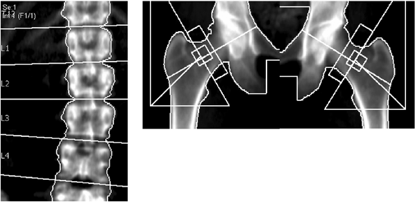
DXA Images
 |
| Region | BMD (g/cm2) | Young-Adult T-Score | Age-Matched Z-Score |
|---|---|---|---|
| L1 | 0.790 | -2.8 | -1.9 |
| L2 | 0.903 | -2.5 | -1.5 |
| L3 | 0.870 | -2.7 | -1.8 |
| L4 | 0.799 | -3.3 | -2.4 |
| L2-L4 | 0.852 | -2.9 | -1.9 |
| Region | BMD (g/cm2) | Young-Adult T-Score | Age-Matched Z-Score |
| Neck | |||
| Left | 0.642 | -2.8 | -1.8 |
| Right | 0.607 | -3.1 | -2.1 |
| Mean | 0.625 | -3.0 | -2.0 |
| Difference 0.034 | -0.3 | 0.3 | |
| Laboratory Findings | |
| Hemoglobin (12–15 mg/dL) | 13 mg/dL |
| Serum creatinine (0.7–1.5 mg/dL) | 0.9 mg/dL |
| ALT (7–35 IU/L) | 30 IU/L |
| 25-OHD (target >30 ng/mL) | 35 ng/mL |
| Intact PTH (10–65 pg/mL) | 54 pg/mL |
| Ionized calcium (1.10–1.30 mmol/L) | 1.15 mmol/L |
| Total serum calcium (8.9–10.3 mg/dL) | 9.0 mg/dL |
| Phosphorus | 3.6 mg/dL |
| 24-hour urine calcium (100–300 mg/24 h) | 435 mg/24 h |
| Bone-specific alkaline phosphatase Premenopausal female: 4.5–16.9 μg/L Postmenopausal female: 7.0–22.4 μg/L Male 25 years and older: 6.5–20.1 μg/L | 20 μg/L |
| 24-hour urine NTx/creatinine Normal adult female Premenopausal: 17–94 nM BCE/mM creatinine Postmenopausal: 26–124 nM BCE/mM creatinine Normal adult male: 21–83 nM BCE/mM creatinine | 18 nM/mM creatinine |
| TSH (0.2–5 UU/L) | 2.18 UU/L |
| ALT, alanine aminotransferase; PTH, parathyroid hormone; NTx, N-telopeptide; BCE, bone collagen equivalents; TSH, thyroid-stimulating hormone. | |
Teaching Points
Dr. Camacho
The patient has osteoporosis based on WHO criteria.
Similar to Case 1, this patient has low Z-scores and appears to have an inappropriate degree of bone loss for her age.
Her metabolic bone work-up showed idiopathic hypercalciuria. Normal urinary calcium excretion is 1.5 to 3 mg/kg/24 hours or 100–300 mg/24 hours. Her vitamin D and parathyroid hormone (PTH) levels are normal, and there is no evidence of excessive intake of calcium. This is likely the reason why she has osteoporosis at a relatively young age. There is a familial tendency for this disease, and her sister and mother should be evaluated as well.
Patients with idiopathic hypercalciuria respond very well to hydrochlorothiazide (HCTZ). She was started on 25 mg every day, and after 3 months her repeat urinary calcium had normalized at 280 mg/24 hours. Alendronate therapy was continued.
Although HCTZ will correct the secondary cause, there is no strong evidence that this medication alone reduces fractures among patients with idiopathic hypercalciuria. Thus, bisphosphonate therapy should be used for these individuals together with the HCTZ.
Urinary N-telopeptide (NTx) is suppressed to premenopausal range because of her prior intake of alendronate.
Case 3
History
A 50-year-old woman was sent for osteoporosis management. She has no history of fractures or height loss. Menopause was at age 47. The patient is still suffering from menopausal symptoms, including hot flashes and vaginal dryness. She is in good general health, maintains an active lifestyle, and does not smoke. She drinks 2 glasses of skim milk per day and eats a few ounces of cheese.
Physical Examination
Physical examination was unremarkable.
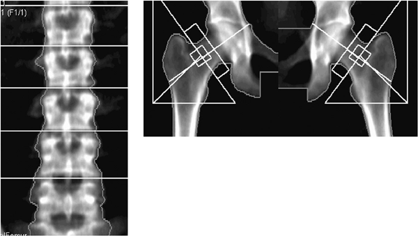
DXA Images
 |
| Region | BMD (g/cm2) | Young-Adult T-Score | Age-Matched Z-Score |
|---|---|---|---|
| L1 | 0.998 | -1.1 | -0.8 |
| L2 | 1.017 | -1.5 | -1.2 |
| L3 | 1.053 | -1.2 | -0.9 |
| L4 | 1.066 | -1.1 | -0.8 |
| L2-L4 | 1.048 | -1.3 | -0.9 |
| Region | BMD (g/cm2) | Young-Adult T-Score | Age-Matched Z-Score |
| Neck | |||
| Left | 0.790 | -2.1 | -1.3 |
| Right | 0.862 | -1.5 | -0.7 |
| Mean | 0.826 | -1.8 | -1.0 |
| Difference | 0.072 | 0.6 | 0.6 |
| Laboratory Findings | |
| Hemoglobin (12–15 mg/dL) | 13.5 mg/dL |
| Serum creatinine (0.7–1.5 mg/dL) | 0.9 mg/dL |
| ALT (7–35 IU/L) | 30 IU/L |
| 25-OHD (target >30 ng/mL) | 35 ng/mL |
| Intact PTH (10–65 pg/mL) | 54 pg/mL |
| Ionized calcium (1.10–1.30 mmol/L) | 1.15 mmol/L |
| Total serum calcium (8.9–10.3 mg/dL) | 9.0 mg/dL |
| Phosphorus | 3.6 mg/dL |
| 24-hour urine calcium (100–300 mg/24 h) | 165 mg/24 h |
| Bone-specific alkaline phosphatase Premenopausal female: 4.5–16.9 μg/L Postmenopausal female: 7.0–22.4 μg/L Male 25 years and older: 6.5–20.1 μg/L | 20 μg/L |
| TSH (0.2–5 UU/L) | 2.18 UU/L |
| ALT, alanine aminotransferase; PTH, parathyroid hormone; TSH, thyroid-stimulating hormone. | |


