Midshaft clavicle fractures in adolescents are common. Recent literature in adults fractures favors open reduction and plate fixation for significantly displaced and/or shortened midshaft clavicle fractures, although whether this applies to adolescents remains debatable. This article reviews the current literature and controversy in the management of displaced adolescent clavicle fractures.
Malunions in adolescent clavicle fractures may not result in major deficits as in adults; however, high level evidence is lacking to support this conclusion.
Malunions in adolescent clavicle fractures may not result in major deficits as in adults; however, high level evidence is lacking to support this conclusion.
Clavicle shaft fractures are among the most common fractures of the upper extremity in adolescents. The incidence of clavicle shaft fractures in the pediatric population is 15% among all upper extremity injuries. Traditionally, clavicle shaft fractures in children and adolescents have been managed nonoperatively, with the exception of open or impending open fractures and fractures associated with floating shoulders or with neurovascular compromise. Most clavicle fractures occur in the shaft along the middle third; this article focuses specifically on clavicle shaft fractures. The optimal treatment of clavicle shaft fractures for older children and adolescents is a topic of major controversy because the literature has shifted more in favor of surgical treatment of displaced clavicle fractures in adults, although whether the research on outcomes in adults is applicable to this younger population remains debatable.
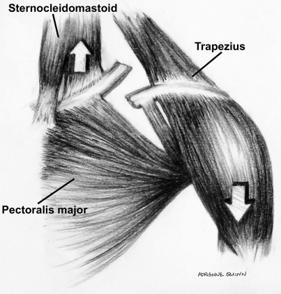
The clavicle is an S-shaped bone when viewed from the axial plane and is a key structure to the shoulder girdle. It has 2 articulations: medially at the sternoclavicular joint and laterally at the acromioclavicular joint. The joint capsules provide stability in both craniocaudal and anteroposterior planes while allowing for rotation occurring with the arc of motion of the shoulder. The clavicle is a key structure of the shoulder suspensory complex, providing ligamentous attachments to the scapula via the conoid and trapezoid coracoclavicular ligaments. In children and younger adolescents, these ligaments attach to a thick periosteal sleeve. Six muscles attach to the clavicle, with the sternocleidomastoid, sternohyoid, and pectoralis major attaching medially; the subclavius muscle attaching along the midportion; and the deltoid and trapezius attaching laterally. Understanding the relative muscular attachments allows for appreciation of direction of displacement of fracture fragments ( Fig. 1 ). The clavicle is also an important structure that protects critical neurovascular structures, including the subclavian artery and vein, and the brachial plexus, especially along the middle third of the clavicle.
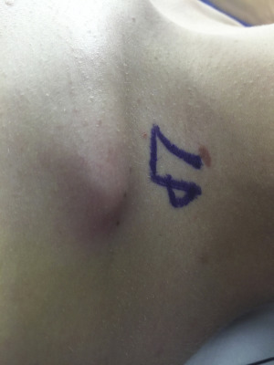
The clavicle shaft is most commonly injured by an axial load to the clavicle transmitted through a fall onto the shoulder. Less common mechanisms of fracture include a direct blow to the clavicle or fall onto an outstretched hand. The middle one-third shaft of the clavicle is the most commonly injured location due to its transitional location leading to a stress riser, where the shape of the clavicle transitions from concave to convex, and from tubular to flat. The direction of displacement depends on both the location of the fracture and the initial force causing the injury. Superior displacement of the medial fragment is common due to the pull of the sternocleidomastoid and can lead to tenting of the skin ( Fig. 2 ) or, in rare instances, open injuries when there is severe displacement. The lateral fragment often displaces inferiorly due to the weight of the shoulder and arm. A thorough neurovascular examination of the injured extremity should be performed to due to the proximity of the subclavian vessels and brachial plexus. Severe displacement or comminution of the clavicle fracture should alert the clinician to a high-energy mechanism, and one should be vigilant in looking for potential associated injuries, including pneumothorax, or fractures of the scapula and ribs. Anteroposterior and 30-degree cephalic tilt radiographs (serendipity view) with the patient in an upright position are sufficient to characterize clavicle shaft fractures. In cases of significant comminution, including radiographs of the contralateral side can aid in assessing the amount of shortening.
Nonoperative treatment generally involves the use of a simple shoulder sling or figure-of-8 brace for approximately 3 to 4 weeks, followed by gradual increase in range of motion. There is an absence of data in the literature supporting that a figure-of-8 brace results in improved outcomes when compared with a shoulder sling, though the figure-of-8 bracing has been associated with more pain and discomfort. Consequently, a shoulder sling has become the preferred nonoperative treatment method. In the adolescent, gradual return to strengthening can be started at 6 weeks, and union should be expected by 12 weeks in most cases. In younger children, fracture healing may be more expeditious.
Surgical technique for plate and screw fixation
For open reduction and internal fixation of clavicle fractures, a superior or anterior plating location can be chosen based on surgeon preference. The patient is positioned in a beach chair or supine position, based on the surgeon’s preference. An approximately 7 to 9 cm horizontal skin incision is made centered along the clavicle. The subcutaneous tissue and platysma is dissected and, if visible, the crossing supraclavicular nerves are identified and preserved. Occasionally, a supraclavicular nerve branch is sacrificed to gain adequate exposure for the open reduction and internal fixation, and it is important to counsel the patient about small regions of potential superior chest wall numbness before surgery. The clavipectoral fascia and surrounding muscle is dissected in a clean plane to allow for eventual closure. The bone is exposed with the periosteal elevator. If 1 large comminuted fragment is present, it is reduced to 1 of the major fragments with a small fracture reduction clamp and a lag screw (generally 2.7 mm or 3.5 mm) is inserted to secure the fragment. The surgeon should be careful to preserve some periosteum on the comminuted fragment to help maintain its viability and facilitate fracture healing. Next, the major bone fragments are directly held by fracture reduction clamps and reduced anatomically. If the fracture configuration allows, it is helpful to either place a 1.6 mm k-wire to hold this reduction or place 1 or more lag screws away from the plane of the intended plate to hold the major fragments together. An anatomically contoured plate is assessed for overall fit along the clavicle, with a goal of at least 3 screws on each of the major fracture fragments. Occasionally, the plates require subtle bending to match the clavicle shape and prevent loss of reduction as the screws are affixed to the plate. The screw holes are drilled with care not to plunge in order not to injure lung or underlying neurovascular structures. Screws are sequentially placed on each side of the fracture, the fixation verified fluoroscopically, and routine irrigation and closure is performed. A sling is used postoperatively for comfort for a maximum of 2 weeks and pendulum exercises to the shoulder are allowed. From 2 to 6 weeks range of motion is gradually progressed. From 6 weeks, gradual strengthening of the shoulder can commence and contact sports can be performed once full radiographic union and strength is achieved.
Related posts:

Stay updated, free articles. Join our Telegram channel

Full access? Get Clinical Tree


