Arthroscopic Suprascapular Nerve Decompression
Eric D. Bava
Sumant G. Krishnan
Suprascapular neuropathy is a less common cause of shoulder pain and dysfunction, but it is now being diagnosed with greater frequency (1, 2). Compression of the suprascapular nerve at either the suprascapular notch or the spinoglenoid notch may occur as an isolated lesion without any other pathology present, or it may occur concomitantly with other pathologies such as a posterior-superior labral tear or rotator cuff tear (1, 3, 4).
INDICATIONS/CONTRAINDICATIONS
Surgical decompression of the suprascapular nerve is considered for patients with suprascapular neuropathy and concomitant shoulder pathology that require operative intervention or for patients with isolated suprascapular neuropathy who have not improved after an initial trial of nonoperative management for usually 3 to 6 months (2, 5). Suprascapular neuropathy may occur in the setting of a massive rotator cuff tear due to a traction injury, (3) or it may be caused by nerve compression from a mass lesion such as a paralabral ganglion cyst associated with a labral tear (4). When suprascapular neuropathy occurs along with a small rotator cuff tear, the supraspinatus and infraspinatus weakness, atrophy, and fatty infiltration seen are greater than would be expected for the size of the cuff tear. Thus, all additional pathologies must be identified and treated in addition to managing the suprascapular nerve.
PREOPERATIVE PLANNING
Patients with suprascapular neuropathy often exhibit pain over the posterior and lateral aspects of the shoulder, as well as weakness of both the supraspinatus and infraspinatus muscles. Physical examination may demonstrate atrophy in either the supraspinatus or the infraspinatus fossa along with decreased strength of the supraspinatus and/or infraspinatus muscles. A careful physical examination can indicate the location of compression along the course of the nerve. Suprascapular nerve dysfunction at the suprascapular notch will exhibit weakness and atrophy of both the supraspinatus and infraspinatus, whereas weakness and atrophy of only the infraspinatus indicate dysfunction more distal along the course of the nerve. Evaluation should also include a complete neurologic examination along with evaluation of the cervical spine. This is important in order to rule out cervical disc disease as a possible cause of neurologic symptoms. A valuable adjunct to physical examination can be a diagnostic injection of 1% lidocaine into the suprascapular notch.
Preoperative studies should include plain radiographs, magnetic resonance imaging, and electromyographic (EMG) testing. Plain radiographs are obtained in order to evaluate additional shoulder pathology such as acromioclavicular and glenohumeral joint arthritis. Other significant findings may include changes about the greater tuberosity and acromion indicative of rotator cuff pathology (Codman changes) and the presence of an os acromiale.
A preoperative MRI is obtained to evaluate the rotator cuff and the glenoid labrum and identify any paralabral cysts or soft-tissue masses that may cause compression of the suprascapular nerve. MRI evaluation is critical in these patients in order to identify any tears of the rotator cuff and also to evaluate for atrophy and fatty infiltration of the cuff musculature. EMG and nerve conduction studies are obtained to confirm and localize the suprascapular nerve compression. This may indicate increased latency, denervation fibrillation potentials, and diminished amplitude in the infraspinatus muscle and, possibly, the supraspinatus muscle depending on the location of nerve compression.
A preoperative MRI is obtained to evaluate the rotator cuff and the glenoid labrum and identify any paralabral cysts or soft-tissue masses that may cause compression of the suprascapular nerve. MRI evaluation is critical in these patients in order to identify any tears of the rotator cuff and also to evaluate for atrophy and fatty infiltration of the cuff musculature. EMG and nerve conduction studies are obtained to confirm and localize the suprascapular nerve compression. This may indicate increased latency, denervation fibrillation potentials, and diminished amplitude in the infraspinatus muscle and, possibly, the supraspinatus muscle depending on the location of nerve compression.
Once the cause and location of suprascapular nerve compression are determined, appropriate management proceeds. If there is no structural compression of the nerve, nonoperative modalities (fluoroscopically guided steroid injection into the suprascapular notch, oral steroid medications, oral nonsteroidal medications, and physiotherapy) are instituted. If these fail after 3 to 6 months with persistent pain and/or weakness, or if there are structural lesions requiring surgical management, surgery should be directed to decompressing the suprascapular nerve at the site of compression.
SURGERY
Surgical Technique
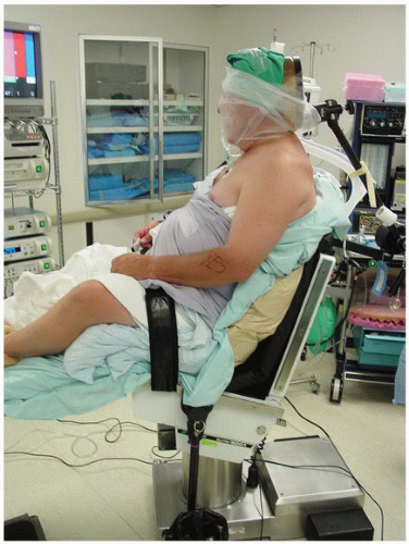
Arthroscopic suprascapular nerve decompression has been described with a number of different techniques (5, 6, 7). Our preference is an anterior approach described previously by the senior author (8) using a modified beach-chair position (Fig. 17-1) and three necessary portals (Fig. 17-2).
The procedure begins with an arthroscopic examination of the glenohumeral joint through a posterior portal and then establishing an anterior portal through the rotator interval utilizing a spinal needle and an outside-in technique. The glenohumeral joint is inspected for any intra-articular pathology, such as tears of the glenoid labrum. If the suprascapular nerve is being compressed at the spinoglenoid notch, it is often due to paralabral ganglion cysts associated with a torn labrum. The suprascapular nerve entrapment can be relieved arthroscopically by decompressing the cyst through the labral tear. An arthroscopic periosteal elevator can be used through the anterior portal, advanced along the junction of the glenoid and labrum, and used to open the tear in the labrum (9). This must be done carefully to avoid injury to the suprascapular nerve that lies 1.8 cm medial to the posterior glenoid rim and 2.9 cm from the superior glenoid rim. Alternatively, an arthroscopic motorized shaver may be used to decompress a paralabral cyst from either the anterior or the posterior portal. The arthroscopic shaver can be used to débride the torn portion of the labrum, the paralabral cyst, and evacuate the fluid from within the cyst cavity.
 FIGURE 17-1
Stay updated, free articles. Join our Telegram channel
Full access? Get Clinical Tree
 Get Clinical Tree app for offline access

|


