Arthroscopic Biceps Tenodesis and Release
Pascal Boileau
Sumant G. Krishnan
Jason Old
Gilles Walch
INDICATIONS/CONTRAINDICATIONS
The long head of the biceps (LHB) is a well-known cause of shoulder pain, due to the multiple possible pathologies of the biceps tendon and its pulley system (1), such as tenosynovitis, prerupture, subluxation, or dislocation of the tendon (2). The majority of degenerative changes in the LHB are associated with pathology of the rotator cuff, as highlighted by Neer (3). The relative importance of these structures in producing symptoms is still uncertain and will probably continue to be so as long as our understanding of the mechanism of pain generation in disease of the rotator cuff remains incomplete. The surgical treatment for disorders of the LHB is removal of the intra-articular portion of the tendon by either tenotomy or tenodesis, which is a common procedure either in isolation or in combination with a rotator cuff repair (1, 4).
Open biceps tenodesis is a common and well-accepted open surgical procedure (1, 4, 5). In 1988, we reported the first results of arthroscopic biceps tenotomy for patients with chronic and significant shoulder pain due to a pathologic biceps tendon in the presence of a massive irreparable rotator cuff tear (6). Because we were familiar with the technique of interference screw fixation for hamstring anterior cruciate ligament reconstruction, we developed a technique for biceps tenodesis using bioabsorbable interference screws. This technique has been used routinely in open surgery since 1996 and arthroscopically since 1997 (7, 8). Arthroscopic techniques using sutures alone or sutures with anchors have been described (9, 10, 11, 12, 13, 14), but several studies have demonstrated the superior biomechanical properties of interference screw fixation (15, 16, 17, 18).
The possible indications for LHB tenodesis or tenotomy include shoulder pain associated with tenosynovitis, prerupture, subluxation, or dislocation of the LHB, or a superior labrum anterior and posterior (SLAP) lesion. Basically, this procedure can be performed in three different clinical situations: (a) in association with arthroscopic rotator cuff repairs; (b) in cases of isolated pathology of the biceps tendon with an intact cuff, especially in young athletes; and (c) in cases of massive, degenerative, and irreparable cuff tears with a pathologic biceps tendon responsible for a painful shoulder. We prefer to perform a tenodesis rather than a simple tenotomy, especially in elderly but active and muscular patients as it avoids the distal retraction and bulging of the muscle at the elbow level (which may be a source of pain during work) and the possible slight decrease in supination strength in the first year after surgery. We do not argue that arthroscopic biceps tenodesis is superior to a simple tenotomy in all patients; it is simply another technical option available for the arthroscopic shoulder surgeon. A frail elderly patient will be a preferable candidate for a biceps tenotomy rather than a tenodesis.
A true pseudoparalytic shoulder due to a massive, irreparable rotator cuff tear is a clear contraindication to an isolated biceps tenotomy or tenodesis, and in such patients, we perform a reverse shoulder arthroplasty to restore active elevation. A thin, fragile, almost-ruptured biceps tendon is the technical limit of arthroscopic biceps tenodesis, and in such patients, we perform a simple tenotomy.
PREOPERATIVE PLANNING
The patient who is a candidate for biceps tenodesis complains of chronic shoulder pain. The dominant side is involved in 80% of the cases. The patient can be elderly and retired, over 65, with or without a history of massive and irreparable cuff tear. The patient can also be younger, around 30 or 40, with a history of an “overused
shoulder” because of his or her profession (mason, painter, gardener) or sports activity (throwing athlete). Onset is progressive in half of the cases; the other half has a history of more or less severe initial traumatic injury. The pain is localized at the anterior part of the shoulder, often radiates in the direction of the lateral part of the elbow, and sometimes can reach the dorsal part of the hand. The pain may occur typically with overhead activity but is often present at rest and may awaken the patient during the night. Most patients have already undergone conservative treatment with rehabilitation programs and corticosteroid injections without any success.
shoulder” because of his or her profession (mason, painter, gardener) or sports activity (throwing athlete). Onset is progressive in half of the cases; the other half has a history of more or less severe initial traumatic injury. The pain is localized at the anterior part of the shoulder, often radiates in the direction of the lateral part of the elbow, and sometimes can reach the dorsal part of the hand. The pain may occur typically with overhead activity but is often present at rest and may awaken the patient during the night. Most patients have already undergone conservative treatment with rehabilitation programs and corticosteroid injections without any success.
Looking at the patient from the back, atrophy of the supra- and infraspinatus is sometimes obvious and leads to the diagnosis of an associated massive cuff tear. Anterior palpation of the shoulder reveals localized pain in the area of the bicipital groove, and more importantly, the pain is recognized by the patient as “my pain.” In dislocations of the LHB, the tenderness is more medial on the lesser tuberosity, and the tendon can sometimes be rolled under the fingers. The cross-arm test is painful with the pain being in the anterior part of the shoulder instead of superior such as in acromioclavicular joint pathology. The impingement signs may be positive, but they are not very specific in our experience. We also find Speed’s test, or the “palm-up” test, useful in diagnosing biceps pathology.
While active and passive motions are usually preserved, loss of motion can alert the astute clinician to several scenarios, including an “hourglass biceps, a dislocated biceps, or a pseudoparalytic shoulder.” A hypertrophied and entrapped “hourglass biceps” often presents with loss of the terminal 10 to 20 degrees of active and passive elevation without loss of rotation. This represents true mechanical locking of the shoulder and should not be confused with a frozen shoulder (19). Patients with a dislocation of the LHB may also present with a very typical clinical picture. Dislocation is often traumatic and almost always associated with a tear of the upper subscapularis. The patient presents with a loss of active elevation above 90 degrees, and it is common to find a limitation of active and passive external rotation because the dislocated biceps tendon tethers the inferior part of the subscapularis. Most importantly, however, when a patient presents with a loss of active elevation, it is crucial to differentiate between true pseudoparalysis of the shoulder and painful loss of elevation. A shoulder with true pseudoparalysis is nonfunctional, exhibiting an ineffective shrug with attempted elevation of the arm, and will not respond to an isolated biceps procedure. A shoulder with painful loss of elevation is functional but active elevation is limited because of pain and often responds well to a biceps tenotomy or tenodesis. Performing the “landing test” can help to differentiate between the two entities (20). The examiner passively places the arm just above the horizontal level (between 90 and 120 degrees). A patient with true pseudoparalysis of the shoulder will not be able to actively maintain the arm in this position and it will fall down despite the patient’s efforts.
For all patients, the preoperative examination should include a radiographic evaluation with a true anteroposterior (AP) view in three rotations and a normalized supraspinatus outlet view. In case of a massive cuff tear involving the infra- and supraspinatus, the acromiohumeral distance, measured on the AP view in neutral rotation, is less than 6 mm. Computed tomography (CT arthrography) or magnetic resonance imaging (MRI) with gadolinium can demonstrate an “unstable” biceps tendon, being either subluxated or dislocated.
SURGERY
The principle of the arthroscopic biceps tenodesis is simple: After biceps tenotomy, the tendon is exteriorized and doubled on a suture and then pulled back into a humeral socket and fixed using a bioabsorbable interference screw. The fixation principle is similar to the interference screw fixation used with success for hamstring anterior cruciate ligament reconstruction of the knee. Doubling the biceps tendon has at least three advantages: (a) it reinforces the strength of the tendon, mitigating damage from the interference screw; (b) it prevents sliding of the tendon after screw insertion (stop-block effect); and (c) it allows optimal tensioning of the biceps muscle since its functional length is not changed.
Patient Positioning
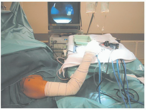
Although the lateral decubitus position can be used, we prefer to perform this technique with the patient in the beach-chair position, under general anesthesia and/or interscalene block. The shoulder should be placed in approximately 30 degrees of flexion, 30 degrees of internal rotation, and 30 degrees of abduction (arthrodesis position), allowing the anterior part of the subacromial bursa to be adequately filled with water in order to have a clear view of the superior part of the bicipital groove. When using the beach-chair position, a classic knee support (horseshoe support) is used with a Mayo stand to allow flexion and extension of the elbow (Fig. 9-1).
Bony Landmarks
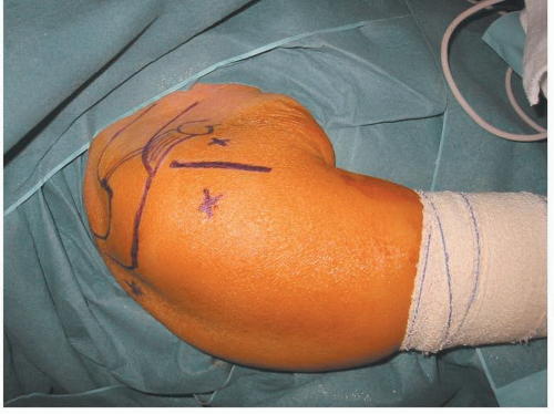
Draw the bony landmarks on the shoulder to identify the spine of the scapula, the acromion, the coracoid process, and the coracoacromial ligament. This procedure requires three arthroscopic portals: The classic posterior portal is created in the “soft spot” 2 cm inferior and 2 cm medial to the posterolateral corner of the acromion; two anterior portals (anteromedial and anterolateral) are created 1.5 cm on each side of the bicipital groove and 3 cm inferior to the anterolateral corner of the acromion (Fig. 9-2). The posterior and anterolateral portals are used for the scope (viewing portals) and the anteromedial portal is used for the instruments (working portal). A pump is helpful to obtain distention of the joint and the bursa, but it is important to maintain low pump pressure (30 mm Hg or less) during the procedure to prevent excessive soft-tissue distention.
 FIGURE 9-2 The posterior portal and the two anterior portals: anteromedial and anterolateral, on each side of the bicipital groove.
Stay updated, free articles. Join our Telegram channel
Full access? Get Clinical Tree
 Get Clinical Tree app for offline access

|


