Arthritis
Osteoarthritis
General Information
In degenerative arthritis or osteoarthritis of the hand, the most commonly involved joints are the distal interphalangeal joints (DIPJ), proximal interphalangeal joints (PIPJ), and thumb carpometacarpal (CMC) joint. The metacarpophalangeal joints (MCPJ) are rarely involved. Marginal osteophytes, producing enlargement of the DIPJ and PIPJ, are known as Heberden’s and Bouchard’s nodes, respectively (Fig. 1).
Arthritis of the Basilar Joint of the Thumb
General Information
Pain and loss of function may result from primary osteoarthritis, posttraumatic arthritis, or rheumatoid arthritis of the carpometacarpal joint of the thumb. Arthritis causes a reduction of pinch and grip strength and necessitates adaptive movements to avoid painful motion of this joint. The joint may narrow, stiffen, and/or subluxate. A resultant adduction deformity of the thumb metacarpal may lead to a secondary hyperextension deformity or other disabling instabilities of the thumb metacarpophalangeal joint. The osteoarthritis may have a genetic basis and be associated with osteoarthritis of other joints. Posttraumatic arthritis may develop from fractures of dislocations of the base of the first metacarpal that result in incongruity of subluxation of the joint. In rheumatoid disease, joint damage results from synovial disease. Osteoarthritis is more common over the age of 40 and approximately three times more common in women than men. Carpal tunnel syndrome occurs with increased frequency in patients with thumb basilar joint arthritis.
Diagnostic Criteria
Clinically, the patient presents with pain with use of the thumb. Swelling or deformity of the basilar joint may be present. There may be a history of a previous
joint injury or rheumatoid arthritis. On physical examination, tenderness and/or dorsoradial subluxation of the thumb carpometacarpal joint is noted. The axial compression-adduction test (Fig. 2) is done by manipulating the thumb with axial compression and adduction. Pain, joint instability, and crepitus are noted at the arthritic carpometacarpal joint.
joint injury or rheumatoid arthritis. On physical examination, tenderness and/or dorsoradial subluxation of the thumb carpometacarpal joint is noted. The axial compression-adduction test (Fig. 2) is done by manipulating the thumb with axial compression and adduction. Pain, joint instability, and crepitus are noted at the arthritic carpometacarpal joint.
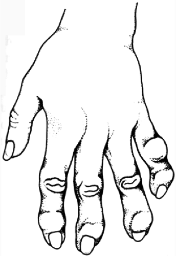 FIG. 1. Osteoarthritis of the hand. |
The axial compression and rotation or grind test (Fig. 3) is performed by compressing and rotating the proximal phalanx and metacarpal of the thumb on the trapezium. Pain is noted at the CMC joint.
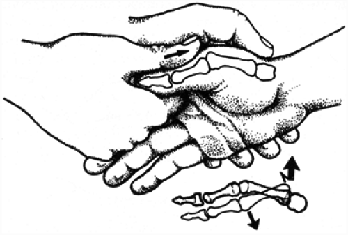 FIG. 2. Axial compression-adduction test of the thumb carpometacarpal joint. |
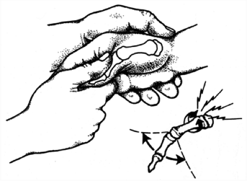 FIG. 3. Axial compression and rotation or grind test of the thumb carpometacarpal joint. |
The relocation test (Fig. 4) is done by placing pressure on the base of a dorsally and radially subluxed thumb metacarpal. Placing pressure in a volar and ulnar direction reduces the CMC joint. Pain is noted at the CMC joint.
Related posts:


Stay updated, free articles. Join our Telegram channel

Full access? Get Clinical Tree


