 Applied Surgical Anatomy of the Approaches to the Hind Part of the Foot
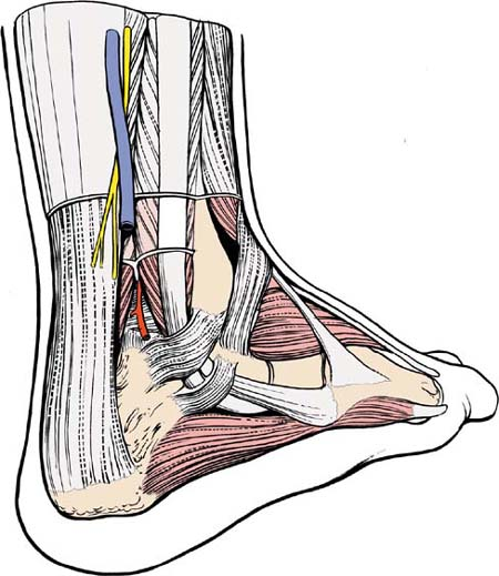
Surgery performed on the hind part of the foot is confined almost exclusively to three joints: the posterior part of the subtalar joint, the talocalcaneonavicular joint, and the calcaneocuboid joint. The anatomy of the approaches is the anatomy of the joints themselves, because they all are superficial structures (see Figs. 25-10 and 26-3).
The key to the anatomy is the tarsal canal, which runs obliquely across the foot, between the talus and the calcaneus. The canal is formed by two grooves, one on the inferior surface of the talus and the other on the superior surface of the calcaneus. The canal separates the talocalcaneonavicular joint from the talocalcaneal joint and acts as a landmark for surgical access to the two joints. At its lateral end, the canal widens considerably into the sinus tarsi.
Stay updated, free articles. Join our Telegram channel

Full access? Get Clinical Tree


