 Anteromedial Approach to the Talar Neck
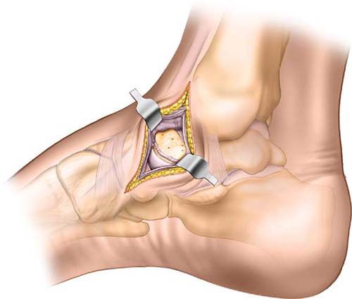
The anteromedial approach to the talar neck offers an excellent view of the medial side of the talar neck. It also permits inspection of the anteromedial portion of the ankle joint, dome of the talus, and talonavicular joint. The anteromedial approach to the talar neck is usually used in conjunction with the anterolateral approach to the talar neck to accurately visualize talar neck fractures. It is generally thought that two incisions are the best approach to deal with this difficult clinical scenario. The two approaches together provide excellent visualization of talar neck fractures for their reduction and fixation.
Position of the Patient
Place the position supine on the operating table (see Fig. 7-1). Place a sandbag beneath the hip on the side undergoing surgery. This will correct the natural external rotation of the leg and place the foot in a neutral position, with the toes pointing skyward. After exsanguination, apply a tourniquet to the mid-thigh.
Landmarks and Incision
Palpate the tendon of the tibialis anterior as it runs over the anteromedial aspect of the ankle. Trace the tendon distally to its insertion on the navicular. Identify the ankle joint by passively flexing and extending the joint. Make an 8-cm-long straight incision on the anteromedial aspect of the ankle. Begin 2 cm proximal to the junction of the medial dome of the talus and distal tibia. Extend the incision distally to follow the medial side of the anterior tibial tendon, ending at the anteromedial border of the navicular (Fig. 14-1). The incision may be extended proximally if access to the medial portion of the ankle is needed. The incision can also be extended distally for access to the medial portion of the midfoot.
Internervous Plane
No internervous plane is used. The approach is safe because the incision cuts down onto bone, which is subcutaneous both proximally and distally.
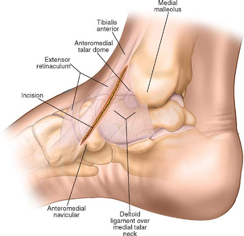 Figure 14-1 Make an 8-cm-long straight incision on the anteromedial aspect of the ankle. Begin 2 cm proximal to the junction of the medial dome of the talus and distal tibia. Extend the incision distally to follow the medial side of the anterior tibial tendon, ending at the anteromedial border of the navicular.
Stay updated, free articles. Join our Telegram channel
Full access? Get Clinical Tree
 Get Clinical Tree app for offline access

|