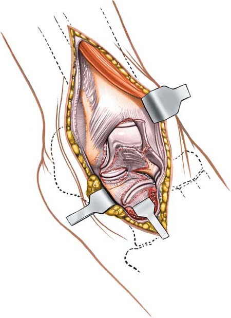
 Anterolateral Approach to the Ankle and Hind Part of the Foot
The full extent of the anterolateral approach to the ankle and hind part of the foot allows exposure not only of the ankle joint but also of the talonavicular, calcaneocuboid, and talocalcaneal joints. The approach is used commonly for ankle fusions, but also can be used for triple arthrodesis and even pantalar arthrodesis. In addition, it is possible to excise the entire talus through this approach, or to reduce it in cases of talar dislocation.
Position of the Patient
Place the patient supine on the operating table; place a large sandbag underneath the affected buttock to rotate the leg internally and bring the lateral malleolus forward. Exsanguinate the limb either by elevating it for 3 to 5 minutes or by applying a soft rubber bandage; then inflate a tourniquet (see Fig. 7-1).
Landmarks and Incision
Landmarks
Palpate the lateral malleolus at the distal subcutaneous end of the fibula.
Palpate the base of the fifth metatarsal, a prominent bony mass on the lateral aspect of the foot.
Incision
Make a 15-cm slightly curved incision on the anterolateral aspect of the ankle. Begin some 5 cm proximal to the ankle joint, 2 cm anterior to the anterior border of the fibula. Curve the incision down, crossing the ankle joint 2 cm medial to the tip of the lateral malleolus, and continue onto the foot, ending some 2 cm medial to the fifth metatarsal base, over the base of the fourth metatarsal (Fig. 9-1).
Internervous Plane
The internervous plane lies between the peroneal muscles (which are supplied by the superficial peroneal nerve) and the extensor muscles (which are supplied by the deep peroneal nerve; see Figs. 25-5 and 25-8).
Superficial Surgical Dissection
Stay updated, free articles. Join our Telegram channel

Full access? Get Clinical Tree


