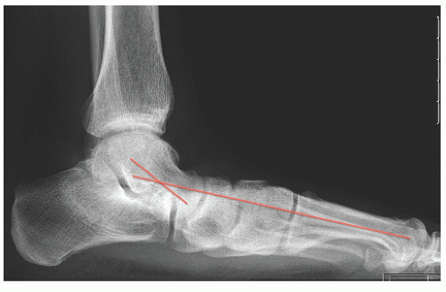
Adult Flatfoot
Steven M. Raikin
Boleslaw Czachor
CLINICAL PRESENTATION
Adult-acquired flatfoot deformity (AAFD) is part of a spectrum of pathologies that are most frequently associated with dysfunction of the posterior tibial tendon (PTT). The PTT is one of the major structures that run behind the medial malleolus and function to support the medial longitudinal arch of the foot. From its origin on the posterior interosseous membrane, tibia, and fibula, the tibialis posterior muscle forms its tendon at the area of the distal metaphyseal flare and then courses around the medial malleolus to insert on the navicular tuberosity, cuneiform, and metatarsals. The tendon functions as a plantarflexor and inverter of the foot.
Tendon failure is most commonly seen in women in their fifth and sixth decades and is frequently associated with obesity. Other factors that can contribute to tendon failure include hypertension, inflammatory arthritis, diabetes, prior foot surgery, or steroid exposure.1
Initial symptoms are variable and correlate with the stage of the disease. Like many other processes associated with aging, failure of the PTT is degenerative in nature. Failure typically occurs immediately at or distal to the medial malleolus in the so-called dysvascular or watershed area of the tendon. This may start as tenosynovitis and can progress to tendinosis with intrasubstance tearing of the tendon and even tendon degenerative rupture.
With regards to symptomatology, as mentioned beforehand, the stages of the disorder help their organization.
Stage 1: Tenosynovitis or tendinosis without foot deformity: The patient may present with medial ankle pain and inflammation that is aggravated by activity, but still have a normal arch.
Stage 2: Tendinosis with flexible arch collapse. This stage is characterized by pain as well as deformity. Pain is usually medially over the PTT, but as the deformity worsens, impingement pain may develop laterally under the tip of the fibular. The deformity involves valgus of the calcaneus and collapse of the arch, but this can be passively corrected (see clinical examination)
Stage 3: The deformity in stage 3 is fixed due to arthrosis of the hindfoot joints. Activity related pain similarly remains the hallmark of this stage and is most commonly seen laterally secondary to subfibular impingement, although medial pain may persist. The differentiation between stage 2 and 3 is made on clinical and radiographic examinations.
Stage 4: Here there is deformity present at the ankle (i.e., the tibiotalar joint) as well as the hindfoot. Due to the severity of the hindfoot valgus, the deltoid ligament becomes increasingly incompetent leading to a lateral tilting of the talus within the ankle mortise. Patients in this stage will usually present with increasing medial ankle joint pain, arthritis, and even potentially stress fractures of the fibular due to the severity of the deformity.2
PHYSICAL FINDINGS
Along with a thorough history that covers possible epidemiologic factors, a thorough physical exam helps support the diagnosis of PTT dysfunction/adult-acquired flat foot. Exam should include inspection for deformity and palpation/range of motion of all of the aforementioned joints (tibiotalar, talonavicular, subtalar, and calcaneocuboid) of bilateral lower extremities. It is critical to examine the patient in both the sitting and standing positions (viewed from behind the patient) to fully appreciate the deformity. Hallmark clinical appearance of the adult-acquired flat foot is the hindfoot in valgus, collapse of the medial longitudinal arch, forefoot abduction (seen as “too many toes” sign) (Fig. 31-1), and Achilles tendon contracture. The examiner can attempt to correct the deformity of the foot passively to differentiate between stage 2 and 3 deformities. A key component of the physical exam
should include the single heel rise maneuver. This can be performed by having the patient elevate up on the toes of the foot that is being examined, while standing on the affected leg. The tested extremity should be in extension at the knee during the maneuver. In a normal foot the patient should be able to get up on the toes and the heel should invert (swing slightly medially toward the medial malleolus). When PTT dysfunction is present, the patient is unable to, or has difficulty and discomfort when attempting to, elevate onto the toes. Additionally, the heel will remain in a valgus position while attempting to perform the heel raise maneuver. Additionally, in the normal foot viewed from behind, the examiner can usually see only the lateral 1 to 2 toes. In the flat foot, multiple toes may be seen, and this is described as the “too many toes” sign. Another clue that could assist with the diagnosis of PTT dysfunction is the posterior tibial edema (PTE) sign as described by DeOrio et al.3 This consists of pitting edema along the course of the PTT. The PTE sign was shown to have 86% sensitivity and 100% specificity for PTT dysfunction when tested against the presence or absence of edema on magnetic resonance imaging (MRI) of the tendon.
should include the single heel rise maneuver. This can be performed by having the patient elevate up on the toes of the foot that is being examined, while standing on the affected leg. The tested extremity should be in extension at the knee during the maneuver. In a normal foot the patient should be able to get up on the toes and the heel should invert (swing slightly medially toward the medial malleolus). When PTT dysfunction is present, the patient is unable to, or has difficulty and discomfort when attempting to, elevate onto the toes. Additionally, the heel will remain in a valgus position while attempting to perform the heel raise maneuver. Additionally, in the normal foot viewed from behind, the examiner can usually see only the lateral 1 to 2 toes. In the flat foot, multiple toes may be seen, and this is described as the “too many toes” sign. Another clue that could assist with the diagnosis of PTT dysfunction is the posterior tibial edema (PTE) sign as described by DeOrio et al.3 This consists of pitting edema along the course of the PTT. The PTE sign was shown to have 86% sensitivity and 100% specificity for PTT dysfunction when tested against the presence or absence of edema on magnetic resonance imaging (MRI) of the tendon.
Stay updated, free articles. Join our Telegram channel

Full access? Get Clinical Tree


