Abstract
Objective
We report the case of a patient who developed paraplegia following a low lumbar epidural steroid injection. Alternative approaches to (or alternative means of) performing transforaminal injections should be considered, in order to avoid devastating neurological complications.
Case report
A 54-year-old man (who had undergone surgery 14 years earlier to cure an L5-S1 slipped disc with right S1 radiculopathy) presented with low back pain (which had begun 6 weeks previously) and left S1 radiculopathy. During a second infiltration of prednisolone acetate, the patient reported feeling a heat sensation in his legs and concomitantly developed facial flushing. Immediately after the injection, the patient developed complete, flaccid T7 ASIA A motor and sensory paraplegia. Three days later, T2 magnetic resonance imaging (MRI) of the spine revealed a spontaneous hypersignal in the conus medullaris and from T6 to T9, suggesting medullary ischemia. Recovery has been slow; after 4 months of treatment in a physical and rehabilitation medicine department, urinary and sensory disorders are still present (T7 ASIA D paraplegia). The patient can walk 200 m unaided. Three months later, the MRI data had not changed.
Discussion
This is a rare case report of paraplegia following low lumbar epidural infiltration via an interlaminar route. The mechanism is not clear. Most of authors suggest that the pathophysiological basis of this type of complication is ischemia caused by accidental interruption of the medullary blood supply. Direct damage to a medullary artery, arterial spasm or corticosteroid-induced occlusion due to undetected intra-arterial injection could result in medullary infarction. This serious incident should prompt us to consider how to avoid further problems in the future. It also raises the issue of providing patients with information on the risks inherent in this type of procedure.
Conclusion
Despite the rarity of this complication, patients should be made aware of its potential occurrence. In the case reported here, the functional prognosis is uncertain.
Résumé
Objectif
L’objectif est de rapporter un cas de paraplégie survenue au décours d’une injection épidurale lombaire basse de corticoïdes pour traiter une lomboradiculalgie S1 et de discuter les mesures à prendre pour éviter cet accident grave à partir de données de physiopathologie et d’une revue de la littérature.
Cas clinique
Il s’agit d’un homme de 54 ans, opéré 14 ans auparavant d’une cure de hernie discale L5-S1, pour une sciatique S1 droite, souffrant d’une lombosciatique gauche, depuis six semaines. Lors de la réalisation d’une seconde infiltration d’acétate de prednisolone, il signale une sensation de chaleur des membres inférieurs, concomitante d’un flush sur le visage. S’installe alors rapidement une paraplégie flasque T7 ASIA A. L’IRM médullaire au troisième jour montre un hypersignal médullaire spontané en T2 au niveau du cône et de T6 à T9, évoquant une myélite ischémique. La récupération est lente. Après quatre mois de prise en charge en médecine physique et de réadaptation (MPR), le patient peut marcher sans aide 200 m. Ils persistent des troubles sensitifs et sphinctériens (paraplégie T7 ASIA D). Les images IRM sont inchangées à trois mois.
Discussion
C’est un cas rare de paraplégie survenant après une infiltration épidurale par voie interépineuse lombaire basse. L’hypothèse physiopathologique généralement retenue est celle d’une ischémie médullaire. Le mécanisme n’est pas clair : injection intravasculaire direct et embolie de microcristaux, atteinte vasculaire (dissection ou spasme d’une artère à visée médullaire), compression vasculaire extrinsèque ou hyperpression localisée dans l’espace épidural ? Cet accident grave doit permettre de définir les moyens pour éviter un nouvel accident. Il pose également le problème de l’information au patient.
Conclusion
Malgré sa grande rareté, cette complication grave mérite d’être connue du patient. Son pronostic fonctionnel est imprévisible.
1
English version
1.1
Introduction
Epidural steroid injection is commonly used in the conservative treatment of degenerative conditions of the lumbar spine (notably for the management of refractory back pain and sciatica). However, this procedure is not inherently benign. Although the complication rate is typically low, paraesthesia, haematoma, epidural abscess, meningitis, arachnoiditis and subdural or arachnoid injections can all occur. Here, we describe another rare complication (acute paraplegia) with two unusual characteristics: (i) epidural steroid injection in the lower lumbar spine (rarely described) and (ii) lesions in two distinct areas of the spine (and not just the conus medullaris). This rare complication raises the issue of patient information and the safety of this procedure, as emphasized recently by the French Agency for Healthcare Product Safety (Afssaps) in a letter sent to rheumatologists and radiologists .
1.2
Case report
A 54-year-old man presented with low back pain and left S1 sciatica which had started 6 weeks previously. There were no neurological impairments. Fourteen years earlier, he had undergone L5-S1 laminectomy to treat right S1 radiculopathy. A clinical examination revealed pain on paravertebral palpation, spinal cord syndrome and a Lasègue (straight-leg) sign at 30°. The neurological examination was normal. A computed tomography (CT) scan revealed L4-L5 and L5-S1 disk damage and right-side L5-S1 paramedian disc herniation. Oral drug treatment was insufficient. An initial lumbar epidural (125 mg of prednisolone acetate) infiltration provided only partial pain relief. One week later, a second infiltration was performed under the same conditions: a 21-gauge intramuscular needle was inserted into median interlaminar region, just above the scar. Aspiration did not recover any cerebrospinal fluid or blood. Again, 125 mg of prednisolone acetate was instilled.
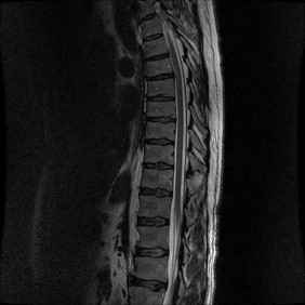
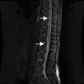
The patient immediately reported feeling a heat sensation in his legs and developed facial flushing. There was no hypotension or faintness. After just a few minutes, the patient developed paralysis of the right lower limb and then acute, flaccid T7 ASIA A paraplegia. Magnetic resonance imaging (MRI) of the thoracolumbar area performed 2 and 7 hours after the onset of symptoms was normal ( Fig. 1 ). Intravenous corticotherapy was introduced for 3 days (120 mg per day on D1 and 240 mg on D2 and D3), followed by 4 days of oral corticotherapy (60 mg per day). Three days later, T2 MRI of the spine revealed a spontaneous hypersignal in the conus medullaris and in the T6 to T9 region, suggesting medullary ischemia ( Fig. 2 ). We did not perform arteriography because of the non-negligible risk of symptom aggravation.


In the following weeks, the patient experienced a slow but steady neurological recovery (beginning in the left leg). Two weeks later, the patient was hospitalized in the physical medicine service. Recovery was slow and at the 4-month follow-up examination, he was still suffering from moderately spastic T7 ASIA D paraplegia. The MRI data had not changed.
The patient was independent in activities of daily living and could walk 200 m unaided. Sphincter dysfunction was still present. A urodynamic analysis ( Fig. 2 ) revealed detrusor hyperactivity with maintenance of bladder volume and the absence of hypocompliance. The patient lacked spontaneous miction and performed four or five intermittent self-catheterizations daily, with no urine leakage. Treatment with oxybutinin (Ditropan ® ) and tamsulosin (Omix ® ) and injection of botulinum toxin into the sphincter did not improve the patient’s urinary status.
1.3
Discussion
Inflammatory phenomena aggravate disc-root compression and primarily occur in the anterior epidural areas. These phenomena can persist and contribute to the continued presence of symptoms in the absence of apparent compression. The powerful anti-inflammatory properties of steroid drugs explain the latter’s use in this indication. Hence, epidural infiltration aims at reaching the anterior epidural areas.
1.3.1
Complications of steroid injection
Complications of epidural steroid injection are generally rare and often mundane: they include the aggravation of low back pain or sciatica, headache, facial flushing and faintness . To date, the Afssaps has recorded just four cases of spinal damage (including the present case) having been declared to France’s Regional Pharmacovigilance Centres following steroid infiltration into the lumbar spine (three cases of paraplegia) and cervical spine (one case of quadriplegia). Two of the cases of paraplegia involved the epidural route (one in L2-L3 and L5-S1 in our patient). The third cases involved left intraforaminal L3-L4 infiltration. In all four cases, neurological motor recovery was only partial.
Our review of the literature identified only eight similar cases . Six occurred after endoscopy-guided or CT-guided foraminal infiltration. Two cases involved secondary paraplegia after lumbar L1-L2 and L4-L5 interlamellar epidural infiltration in the absence of endoscopic guidance. Although no MRI data were available for the latter case, a compressive aetiology was ruled out. All the patients (five men and three women) were aged over 40. Five had a history of lumbar surgery.
Quintero et al. reviewed a number of publications. In every case, the patient described violent pains and extremely rapid-onset neurological impairment. The MRI demonstrated ischemia of the conus medullaris.
Our case report is the second case to have arisen after a low lumbar epidural interlaminar infiltration. The patient felt a heat sensation (rather than pain) and facial flushing before the impairment but after the infiltration. There was also a lag in terms of the MRI data (normal images in the first few hours). In contrast, the ischemia affected two areas: the conus medullaris and T6 to T9.
Intravenous corticotherapy is sometimes reported; its influence on the functional prognosis has not been determined. Although the neurological impairment does not improve in most cases, some partial recoveries have been described. Furthermore, the delayed appearance of MRI anomalies (more than 6 hours later) and/or the subsequent normalization of the MRI signal appear to be associated with a better functional prognosis.
One case deserves special attention . The authors did not report ischemia but observed neurological impairment which worsened over 48 hours. The infiltration had been complicated by puncture of the thecal sac. This case will not be considered in our discussion, since it constitutes another complication of this procedure.
So, which preventive measures can be implemented? In fact, they depend on the pathophysiological mechanism.
1.3.2
Pathophysiological mechanisms
Several hypotheses (linked to either vascular damage or steroid drugs or both) have been proposed. The goal is to understand and explain the causes and then come up with guidelines for avoiding the inherent risks of this procedure.
1.3.2.1
Vascular damage
The neurologic impairment’s speed of onset argues in favour of the presence of a vascular injury. Medullary ischemia would explain the medullary hypersignals in MRI. However, the mechanism of this ischemia is not clear.
Below T8, the arteria radicularis magna (the major anterior radicular artery, also known as the artery of Adamkiewicz) constitutes the major blood supply to the anterior spinal artery . The artery of Adamkiewicz arises between T9 and L2 (from the left, in 85% of cases) in 85% of individuals and at the upper lumbar level in 10% of cases . However, its origin is highly variable; in a small proportion of subjects, the artery of Adamkiewicz may arise from near the upper vertebrae in the lumbar spine (or lower, near L3, L4 or L5) , the thoracic spine (T5 to T8, with a lumbar or sacral supply in 10 to 15% of cases) and, more rarely, from even as low as S1.
Low lumbar infiltration can damage an artery. The artery of Adamkiewicz is an enlarged, radicullomedullary artery which anastomoses with the anterior spinal artery; it extends as far as the lower part of the medulla spinalis and continues as a slender branchlet over the filum terminale. The anterior and posterior branches of the artery in the lumbar enlargement come together at the top of the conus medullaris (near L1 and L2) to form the latter’s anastomosing handle. Below the conus medullaris, the roots of the cauda equina (formed by the L2 to S5 roots) each receive a lower radiculomedullary artery; some terminate at the root, whereas others constitute a lower medullary supply which reaches the terminal conus medullaris (forming part of the latter’s anastomosing handle). This illustrates the vascular complexity of the cauda equina’s roots and the fragility of that of the conus medullaris (a single-end artery with practically no substitute). Hence, one can easily envisage that damage to the latter blood supply may cause a ventral medullary infarct and, as a result, complete paraplegia.
Compression by the injected volume is a poor hypothesis, given the lack of subsequent neurological recovery and the MRI images. Indeed, the liquid spreads out quickly or is quickly resorbed.
Might the needle have caused direct injury to an abnormally low, dominant radiculomeddulary artery (a known anatomical variant)? In support of this hypothesis, Sullivan et al. noted that the risk of intravascular uptake for foraminal infiltration is 10.8%. Contrast agent uptake by a medullary artery is frequently observed. However, preinjection aspiration failed to produce blood flashback in 74% of cases that proved to be intravascular. Hence, aspiration has very poor sensibility ; it should be performed but does not rule out intravascular injection.
One can suppose that the existence of associated vascular abnormalities increases the likelihood of vessel presence in the foramen. We cannot say whether or not our patient has a malformation which might make this type of complication more likely, such as reported by Quintero et al. (angiomatous malformation of T9). In the latter study, no lesions were seen in MRI but an arteriography was not performed.
So, what type of mechanism might be involved? Glaser and Falco suggested (by analogy with other arterial beds) that injury could result from an arterial spasm and/or the development of an intimal flap caused by a direct needle lesion. Both of these mechanisms could lead to stasis flow, clot formation, and hypoperfusion, as seen in coronary lesions . The diagnosis of this type of damage is problematic; arteriography could perhaps have provided more information in the present case.
Our case report describes a rare case of paraplegia following low spinal infiltration. The MRI images in two different areas of the spine suggested that embolization was more likely than a spasm or a direct arterial damage.
1.3.2.2
Causal links with corticosteroids
The Afssaps has emphasized that:
- •
only cortivazol and prednisolone have received regulatory approval for epidural infiltration. Prednisolone is also authorized by intradural injection in the treatment of epidural-refractory sciatica and back pain;
- •
other glucocorticoids (such as triamcinolone and methylprednisolone acetate) are only indicated for intra-articular, peri-articular and loco dolenti injections.
The latter two drugs were injected in five of the nine cases of medullary ischemia described in the literature.
Corticosteroid formulations contain microcrystals which may potentially occlude arterioles measuring 5–10 μm in diameter (such as the medullary vessels). To test this hypothesis, five commonly used preparations (methylprednisolone, triamcinolone, betamethasone sodium phosphate/betamethasone acetate and dexamethasone) were subjected to microscopic analysis. An unopened vial was shaken vigorously. The measurements were recorded and exported to a spreadsheet program. Measurements were sorted according to the particle size. We calculated the proportion of each size class as a percentage of the total number of particles. Aggregates (generally measuring between 1 and 10 μm) were found in each preparation. Some particles exceeded 50 μm in diameter (8.57%, 3.7%, 1.14%, 3.7% and 0% for the five drugs, respectively). The methylprednisolone and the triamcinolone even contained 100 μm particles. The diameter of the medullary arterioles is estimated to be between 10 and 15 microns, with the small medullary arteries measuring around 50 microns. Hence, embolization of a medullary artery by this type of particle could explain the damage described in the present case report. Although solutions of these steroid compounds are valuable because of their long-acting properties (from 36 to 72 hours), the risk may be greater than for suspensions .
Most of the reported cases involved steroid solutions but the present case and those reported by Quintero et al. and Lenoir et al. involved suspensions (a combination of three different compounds in Meyer et al. ).
Benzyl alcohol (the preservative agent in tiamcinolone) may have neurotoxic activity . The damage described in the various case reports does not argue in favour of such a phenomenon but these data suggest that caution should be taken when using these compounds.
1.3.2.3
Other factors
Although a history of spinal surgery is frequently described, it is not clear whether this factor has a role in the occurrence of paraplegia after injection .
The number of injections prior to an incident is often poorly reported in the literature. Some of the patients displayed the complication after one injection, whereas two or four injections were performed in other cases.
1.3.2.4
Clinical practice recommendations
Epidural infiltration is recommended when the patient presents subacute sciatica (regardless of the presence or absence of low back pain). The procedure is more rarely recommended for lumbago. Oral medication and physiotherapy are the recommended front-line treatments. Efficacy is better for recent-onset pain (1 to 60 days) than chronic pain (12 weeks to 1 year) .
There is no consensus on clinical practice recommendations for preventing these complications. Guidelines were initially proposed by Tiso et al. for cervical foraminal injections and were modified by Quintero et al. for lumbar foraminal infiltrations:
- •
prefer a blunt-tipped needle, to reduce the risk of direct vessel lesions;
- •
prefer a large-diameter needle (less than 22 G), to reduce the risk of cannulating a vessel;
- •
perform a preinjection aspiration (it must be negative for cerebrospinal fluid and blood);
- •
use the steroid with the fewest large particles (cortivazol and prednisolone have regulatory approval in France);
- •
the use of contrast agent adds a margin safety by ensuring neural spreading and the lack of vascular uptake (check the position after handling the needle);
- •
use microbore extension tubing to minimize needle handling after a confirmed placement. Medications are injected through the catheter, instead of through the needle. Multiple-level injections require the use of multiple microbore extensions;
- •
inject a local anaesthetic “test” (e.g. adrenaline-free lidocaine) and allow time to pass (time not defined) to detect the occurrence of neurological signs before injection of the corticosteroid (hence the importance of not using sedatives);
- •
inject the corticosteroid slowly. Check regularly for the absence of blood in the aspirate.
These clinical practice recommendations are empirical and result from previously described observations. The various ways of performing the infiltrations have never been compared in a clinical study. Meyer et al. pointed out that some spinal injuries have been described after injection of anaesthetics, whereas this type of drug is given before corticosteroids. Adrenaline-containing formulations must not be used.
The sacrococcygeal route may be of value: there is less risk of damage to the blood vessels in general and major anterior radicular artery in particular. This procedure is not mentioned in the indications.
1.4
Conclusion
The benefits of the epidural injection must always be weighed up against the risk of complications – the most serious of which is neurological injury. Despite its rarity, practitioners must be made more aware of the risk of paraplegia following medullary ischemia.
Stay updated, free articles. Join our Telegram channel

Full access? Get Clinical Tree


